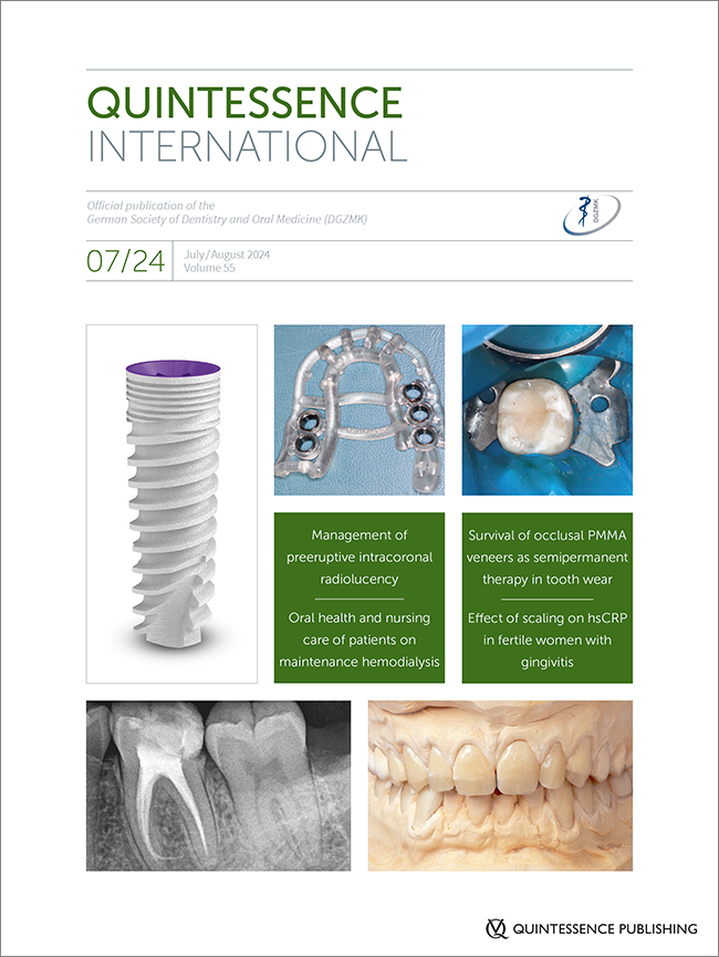
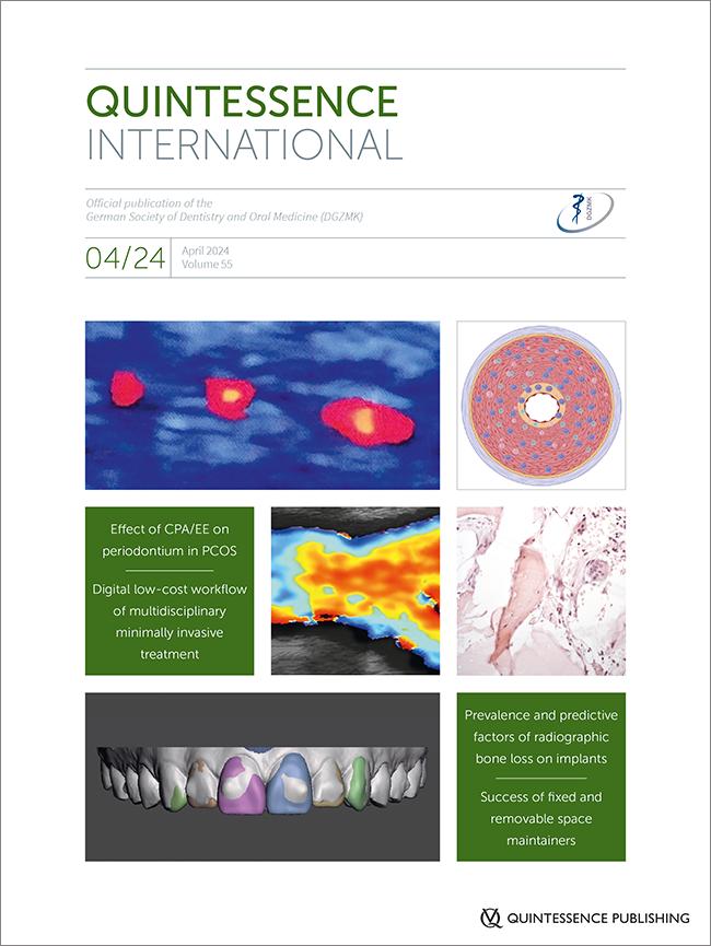
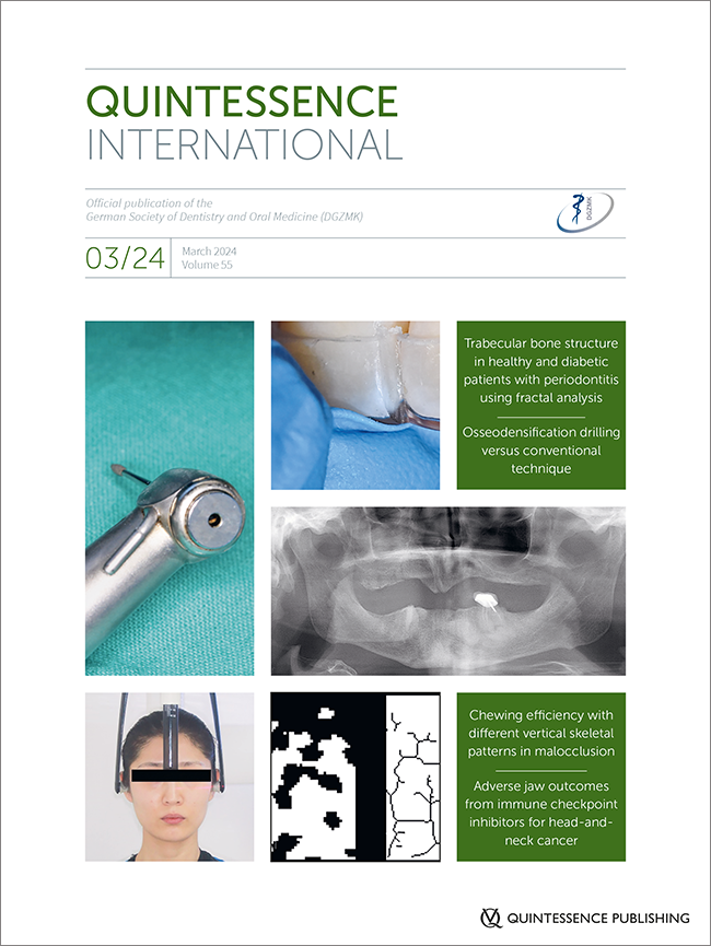
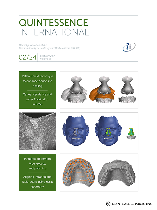
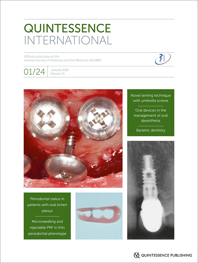
DOI: 10.3290/j.qi.b5721675, PubMed ID (PMID): 39219373Pages 516-517, Language: EnglishGajendra, Sangeeta / Zusman, Shlomo PaulEditorialDOI: 10.3290/j.qi.b5517911, PubMed ID (PMID): 38934773Pages 518-529, Language: EnglishAhlers, M. Oliver / Roehl, Jakob C. / Jakstat, Holger A. / Kielbassa, Andrej M.Objectives: To evaluate the survival rate of minimally invasive semipermanent occlusal polymethyl methacrylate (PMMA) onlays/veneers in previous temporomandibular disorder (TMD) patients with severe tooth wear and with a loss of vertical dimension after up to 7 years. Method and materials: This case series was designed as a follow-up evaluation with consecutive patient recruitment. All patients bearing the indication for this kind of rehabilitation were treated by the same clinician using the same adhesive methodology. The study included 22 patients (3 men/19 women), with a mean ± SD age of 50.7 ± 11.6 years. Controls followed within the first 4 weeks (and subsequently as required). Failure criteria included damage by fracture, chipping, and retention loss. Survival rates were determined based on the Kaplan–Meier analysis. Results: 328 semipermanent occlusal/incisal veneers were included (142 maxillary/186 mandibular teeth). Almost 80% of the restorations were in place and in function when starting the follow-up treatment after 180 days; failures predominantly occurred within the first 3 to 6 months but proved reparable. Depending on the patients’ priorities, scheduled replacements followed successively, and more than 65% did not show repair or any renewal needs for more than 360 days. Conclusion: Within the limitations of this study the survival rates of occlusal veneers made of PMMA were sufficiently high to allow for consecutive treatment of the respective teeth by means of permanent restorations while preserving the restored vertical dimension. In patients with severe tooth wear and a TMD history, semipermanent restorative therapy with occlusal PMMA onlays/veneers would seem a noteworthy option.
Keywords: attrition, case series, occlusal onlays, occlusal veneers, open bite, oral rehabilitation, polymethyl methacrylate, temporomandibular disorder (TMD), tooth wear, vertical dimension, wax-up
DOI: 10.3290/j.qi.b5465309, PubMed ID (PMID): 38874210Pages 530-538, Language: EnglishKrikeli, Eleni / Lambrianidis, Theodoros / Molyvdas, Ioannis / Mikrogeorgis, GeorgiosObjectives: The purpose of the present study was the radiographic evaluation of endodontically treated teeth presenting periapical radiolucency and unintentional overfilling with gutta-percha or sealer on treatment outcome and persistence of the extruded materials. Method and materials: After assessment using periapical index (PAI), 202 roots filled with gutta-percha and zinc oxide–eugenol sealer (Roth 811, Roth International), exhibiting unintentional overfilling and periapical radiolucency were selected. All cases had at least 1 year of follow-up. Type of extruded material, periapical status, and removal/persistence of the extruded material were evaluated by two independent observers. Data were statistically analyzed using logistic and linear regression analysis. Results: Tooth location (P .001), follow-up period (P .001), and type of extruded material (P = .004) significantly influenced treatment outcomes. Specifically, posterior roots exhibited better outcomes compared to anterior, and cases with overfilling of sealer showed superior healing potential compared to those with gutta-percha overfilling. Additionally, longer recall periods were associated with improved treatment success. The type of extruded material (P .001) and follow-up period (P .001) significantly affected the presence of extruded material in the follow-up radiograph. The persistence of extruded material was greater when gutta-percha was extruded, and extruded materials were less detected when the follow-up period was longer. Conclusion: Teeth with periapical radiolucency and unintentional overfilling require longer follow-up intervals for effective monitoring of healing. Treatment outcome was associated with the type of extruded materials used in the present study. The persistence of those materials in the periapex did not affect healing.
Keywords: endodontically treated teeth, overfilling, periapical radiolucency, radiographic evaluation, treatment outcome
DOI: 10.3290/j.qi.b5437505, PubMed ID (PMID): 38847139Pages 540-546, Language: EnglishVerma, Richa / Tewari, Shikha / Anand, DeeptiObjective: Varying levels of sex hormones across the menstrual cycle in young systemically healthy females may alter tissue responses to plaque, resulting in increased gingival inflammation. Also, higher severity and prevalence of gingivitis has been demonstrated in adult women than men, attributed to hormonal changes. Further, it has been reported that gingivitis raises the levels of systemic inflammatory markers such as C-reactive protein. This interventional trial aimed to evaluate the effect of supragingival scaling on serum high-sensitivity C-reactive protein (hsCRP) levels along with periodontal parameters in systemically healthy women of reproductive age with natural gingivitis. Method and materials: In total, 57 women of reproductive age were enrolled into two groups. The test group (n = 30) comprised systemically healthy women with gingivitis who received supragingival scaling. The control group (n = 27) included systemically and periodontally healthy women. Periodontal parameters (Gingival Index, Plaque Index, pocket probing depth, bleeding on probing) and serum hsCRP levels were recorded at baseline for both the groups. Follow-up of test group participants was done at 3 and 6 months. Results: Serum hsCRP and periodontal parameters were significantly higher in the test group than in the control group at baseline, and this decreased significantly after treatment in the test group at the 6-month follow-up (P ≤ .05). Gingival Index, bleeding on probing, and hsCRP in the test group at 6 months were reduced to the baseline levels of systemically and periodontally healthy women. Conclusion: Treatment of gingival inflammation can help in lowering the systemic and local inflammation to the levels of systemically and periodontally healthy women.
Keywords: C-reactive protein, dental scaling, gingival bleeding on probing, gingivitis, inflammation
DOI: 10.3290/j.qi.b5414733, PubMed ID (PMID): 38818638Pages 548-558, Language: EnglishHalabi, Dima / Slutzkey, Gil / Meir, Haya / Sebaoun, Alon / Beitlitum, IlanObjective: To evaluate the survival of fully guided implants placed with a hollow tooth-supported computerized surgical guide (TSSG). Method and materials: This retrospective study included 94 patients who underwent implant placement using freehand or TSSG by the same operator between 2015 and 2020. Early implant failures occurring within 1-year post-rehabilitation were assessed. Results: In the study, two types of implants were placed using two different techniques: TSSG and freehand. The TSSG group consisted of 84 S implants and 100 LP implants, and the freehand group included 90 S implants and 94 LP implants. The results showed that more implants survived when placed freehand compared to TSSG (181 [98.4%] vs 172 [93.5%], respectively, P .05). The only significant factor affecting the success rate was the type of implant, with LP implants having a higher survival rate in the TSSG group (P .05). Conclusion: Surgeons should consider the impact of implant type on survival rates when utilizing the TSSG system.
Keywords: computer-guided implant placement, dental implant, freehand placement, smoking, survival rate
DOI: 10.3290/j.qi.b5223635, PubMed ID (PMID): 38634627Pages 560-568, Language: EnglishAhmed, Eilaf E. A. / Vielhauer, Annina / Splieth, Christian H. / Schmoeckel, Julian / Mourad, Mhd SaidPreeruptive intracoronal radiolucency (PEIR) is a rare dental anomaly often incidentally detected during routine radiographic examinations. This condition manifests as a radiolucent lesion beneath the enamel–dentin junction of unerupted teeth, particularly in mandibular molars, posing diagnostic and management challenges due to its asymptomatic nature. The treatment of PEIR depends on the extent of the lesion and the degree of pulp involvement. Case series: This case series reports on four patients with progressive PEIR. In Cases 1 and 2, lesions were incidentally discovered in panoramic radiographs during orthodontic planning (mandibular permanent second molars), and additional surgical exposure to access the lesion was required as teeth were only partially erupted. Interestingly, in Case 3, the PEIR was not visible in earlier radiographs though the crown of the tooth was already mineralized (mandibular permanent second molar). For Case 4, the tooth presented with symptoms of reversible pulpitis (mandibular permanent first molar). All lesions were treated with indirect pulp capping using biocompat-ible material. The patients were followed up for a period of up to 8 years to evaluate treatment success. Indirect pulp capping and restorations were found to be successful in all four cases in the last follow-up: 1 year (Case 2), 1.4 years (Case 4), 1.5 years (Case 1), and 8 years (Case 3). Conclusion: This case series demonstrates the effectiveness of early intervention via surgical exposure and indirect pulp capping and restoration for managing severe cases of PEIR. However, further research with larger samples and long follow-up is necessary.
Keywords: preeruptive intracoronal radiolucency, preeruptive intracoronal resorption, pulp capping, radiolucent lesion, unerupted teeth
DOI: 10.3290/j.qi.b5223619, PubMed ID (PMID): 38634626Pages 570-578, Language: EnglishSayed Taha, Aisha M. / Almahdi, Wael H. / Alhamad, Nada A.Objectives: The frenum is a mucous membrane fold that attaches the lip and the cheek to the alveolar mucosa, the gingiva, and the underlying periosteum. Frenectomy is the surgical removal of the whole frenum, including the area connected to the bones. The purpose of this study was to compare the healing period and postsurgical pain experienced by patients operated with diode and Er:YAG lasers. Method and materials: Twenty referred patients requiring excision of the abnormal upper labial frenum were included in the study. Patients were randomly assigned into two groups: diode group (810 nm, 2W, continuous emission, initiated tip) and Er:YAG group (2,940 nm, 2W, 200 mJ, 10 Hz). Both lasers were applied in contact mode. Postoperative pain was assessed using a numerical rating scale at 3 hours postoperatively and every day during the first postoperative week. The epithelialization process of the wound surface was evaluated using hydrogen peroxide solution applied to the wound on postoperative days 7, 14, 30, 60, and 90. Results: The results showed the mean values of Pain Index after 3 hours (diode group 2.1 ± 2.0, Er:YAG group 2.6 ± 1.4), day 1 (diode group 1.1 ± 1.1, Er:YAG group 1.9 ± 1.4), and day 2 (diode group 0.0 ± 0.0, Er:YAG group 0.9 ± 1.1), with no significant difference after 3 to 7 days (P = 1.00). For the Healing Index there was a significant difference between the diode group and the Er:YAG group (7 days, P = .029; 14 days, P = .001), with no significant difference after 30/60/90 days (P = 1.00). Conclusions: The Er:YAG laser had better clinical results in healing wounds, whereas the diode laser resulted in better decreasing pain levels after frenectomy during the follow-up periods.
Keywords: YAG laser, frenectomy, papilla penetrating, papillary
DOI: 10.3290/j.qi.b5342511, PubMed ID (PMID): 38757949Pages 580-588, Language: EnglishZhang, Qinglai / Zhang, Yue / Lin, Lili / Meng, Fei / Jia, MengObjective: To determine the oral health status of patients on maintenance hemodialysis and to identify the factors influencing their oral health. Method and materials: This observational study included 1,186 patients with chronic kidney disease who received maintenance hemodialysis across 33 hospitals in China. The patients were recruited for a questionnaire survey between April and August 2023 at Beijing Chaoyang Hospital using stratified sampling. Data collection tools included the General Information Questionnaire for Maintenance Hemodialysis Patients, the Oral Health Assessment Tool, the Pittsburgh Sleep Quality Index, and the Hospital Anxiety and Depression Scale. Spearman rank correlation coefficients were used to assess the relationships between the oral health of patients on maintenance hemodialysis and continuous variables such as sleep quality and emotional status. Multiple linear regression analysis was employed to explore the relationship between oral health and various variables. Results: The oral health scores of the patients ranged from 8 to 22, with a mean score of 12.54 ± 2.63. The final model of the multiple linear regression analysis indicated a goodness of fit of 22.19%. Independent factors affecting the oral health of patients included smoking, the proportion of medical expenses, water consumption, sleep quality, and anxiety scores (all P .05). High levels of smoking, substantial medical expenses, poor sleep quality, and elevated anxiety scores were risk factors for poor oral health (all P .05). Adequate daily water intake served as a protective factor for oral health (P .05). Conclusion: This study proposes targeted interventions to enhance the management and improvement of oral health in patients on hemodialysis, aiming to provide highly personalized and effective oral health care. These interventions are expected to improve oral health outcomes in future clinical practice.
Keywords: influencing factors, maintenance hemodialysis, nursing care, oral health
DOI: 10.3290/j.qi.b5566159, PubMed ID (PMID): 38985438Pages 590-600, Language: EnglishDegirmenci, Kubra / Yalın, MelikeObjectives: This study aimed to evaluate the effect of the clinical removal of fixed partial dentures on oral health-related quality of life and the anxiety values of individuals and to determine the clinical factors of high anxiety levels. Method and materials: In total, 300 participants were included in the study. Six different reasons for the clinical removal of fixed partial dentures (oral examination, denture renewal, endodontic treatment, tooth extraction, periodontal treatment, and composite filling restoration) were defined. The United Kingdom Oral Health-Related Quality-of-Life Measure (OHRQoL-UK), the Modified Dental Anxiety Scale (MDAS), and the Spielberger State-Trait Anxiety Inventory- State (STAI-S) and Trait (STAI-T) were answered. The reason groups were compared using one-way analyses of variance. Binary logistic regression analyses were performed to evaluate the risk factors for high anxiety. Results: There was no significant difference in OHRQoL-UK scores (P = .279) among the reason groups, but there were significant differences in MDAS, STAI-S, and STAI-T scores (P = .004, P .001, P = .018, respectively) among the reason groups. Endodontic treatment, tooth extraction, and sex were determined to be risk factors, considering the anxiety scales. Conclusions: Females are 2.2 times more likely to have trait anxiety than men. Although the effect of the reason for the clinical removal of fixed partial dentures on oral health-related quality of life was similar among the groups, it is concluded that endodontic treatment and tooth extraction reasons for the clinical removal of fixed partial dentures could be risk factors for high anxiety regardless of fixed partial denture usage time.
Keywords: anxiety, dental anxiety, fixed partial denture, oral health-related quality of life, Spielberger State-Trait Anxiety Inventory
