Various cookies are used on our website: We use technically necessary cookies for the purpose of enabling functions such as login or a shopping cart. We use optional cookies for marketing and optimization purposes, in particular to place relevant and interesting ads for you on Meta's platforms (Facebook, Instagram). You can refuse optional cookies. More information on data collection and processing can be found in our privacy policy.
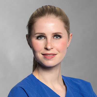
2013 – 2018 Studium der Zahnmedizin an der Albert-Ludwigs-Universität Freiburg. Seit 2018 Zahnärztin und wissenschaftliche Mitarbeiterin in der Abteilung für Zahnärztliche Prothetik, Alterszahnmedizin und Funktionslehre, Charité Berlin. 2022 Abschluss der Promotion. 2023 Ernennung zur qualifiziert fortgebildeten Spezialistin der Deutsche Gesellschaft für Prothetische Zahnmedizin und Biomaterialien e.V. (DGPro).
Events
37. Berliner Zahnärztetag
KEIN MUT ZUR LÜCKE! Prothetik und Ästhetik von A bis Z21. Apr 2023 — 22. Apr 2023Estrel Convention Center
Speakers: Giuseppe Allais, Sven-Marcus Beschnidt, Florian Beuer, Maria Bruhnke, Vincent Fehmer, Petra Gierthmühlen, Christian Haase, Arndt Happe, Marie-Elise Jennes, Christopher Köttgen, Ina Köttgen, Andreas Kunz, Jan Kurtz-Hoffmann, Otto Prandtner, Irena Sailer, Guido Sterzenbach, Dana Weigel
Quintessenz Verlags-GmbH
This author's journal articles
Quintessence International, 6/2022
DOI: 10.3290/j.qi.b2793257, PubMed ID (PMID): 35274516Pages 534-545, Language: EnglishJennes, Marie-Elise / Hey, Jeremias / Bartzela, Theodosia N. / Mang de la Rosa, Maria R.
The treatment management of patients with hemifacial microsomia (HM) includes both surgical and nonsurgical approaches and depends primarily on the degree of deformity of the facial and skeletal structures. In this context, the combined efforts of the maxillofacial surgeon, the orthodontist, and the prosthodontist are essential for a satisfactory functional and esthetic outcome.
Case presentation: A 31-year-old man presented with a chief complaint of facial asymmetry. The patient had been diagnosed with HM on the right side, with severe external ear deformity, and hypoplasia of the facial muscles and the zygomatic bone. The intraoral examination showed a Class I molar and canine relationship with a reduced horizontal overlap and an occlusal plane canting. The maxillary anterior teeth were severely worn due to traumatic occlusion. Orthodontic treatment in conjunction with combined orthognathic surgery was planned to address the facial asymmetry. Ramus distraction osteogenesis was carried out, followed by conventional presurgical orthodontic treatment. The treatment was completed by prosthetic rehabilitation for the reconstruction of the maxillary teeth and fine occlusal adjustment.
Conclusion: The cooperation between the orthodontist, surgeon, and prosthodontist becomes indispensable when treating complex cases of HM. An interdisciplinary approach should be adopted from the start of treatment, promoting integrated customized care.
Keywords: functional rehabilitation, hemifacial microsomia, interdisciplinary treatment, orthodontics, prosthodontics
Aim: The present narrative review aims to provide an overview of the in vivo accuracy of full-arch scans performed with currently used intraoral devices and to compare different methods for the determination of in vivo accuracy.
Materials and methods: An electronic search was performed with the PubMed, Cochrane Central Register of Controlled Trials (CENTRAL), and Web of Science databases, including articles from 2015 to 2020. Specific search strategies were developed for each platform.
Results: The final search resulted in five published articles. The mean values of trueness and precision of the examined scanners ranged from 12.9 to 80.01 µm for trueness and from 42.9 to 86.0 µm for precision in full-arch dentition. Not all studies evaluated both trueness and precision. Furthermore, the methods and references for determining the in vivo accuracy proved to be very different.
Conclusion: To date, no scientific consensus on the required accuracy of in vivo full-arch scans has been reached and published. Due to the small number of studies on the in vivo accuracy of full-arch scans, further studies should be conducted in this area. In addition, a concept for a valid reference that can be scanned both extraorally and intraorally should be developed.
Keywords: CAD/CAM, accuracy, in vivo accuracy, intraoral scanner, optical impressions, digital impression, digital dentistry