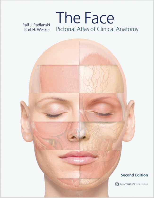
The Face
Pictorial Atlas of Clinical Anatomy
2nd revised Edition 2015
Book
Hardcover, 24 x 30 cm, 368 pages, 380 illustrations
Language: English
Categories: Anatomy, Facial Esthetics, Oral/Maxillofacial Surgery
ISBN 978-1-85097-289-1
QP Deutschland
Buy this product from the following publishers:
Buy at QP Deutschland Buy at QP United Kingdom Buy at QP USAFor the first time, the highly complex topographic-anatomical relationships of facial anatomy are depicted layer by layer using extremely detailed anatomical illustrations with a three-dimensional aspect. Important landmarks, anatomical details, and clinically relevant constellations of hard and soft tissues, as well as of nerves and blood vessels, have been detailed. Another important feature is that the point of view is maintained throughout while moving through the different layers of preparation. While the accompanying text and figure captions highlight specific issues, the images remain in the foreground. The elaborate illustrations are based mainly on live anatomy and corresponding images obtained from magnetic resonance imaging, with some support from anatomical preparations.
Contents
Chapter 1. The Face
• Introduction
• The face in anterior view
• The face in lateral view
• The head in vertical view
• The head in dorsal view
• The neck
• Facial expression
• The facial skeleton
• Sectional anatomy
• Schematic representations of pathways in the face
Chapter 2. The Orbital Region
• Surface topography of the orbital region
• Preseptal muscles and fat layers
• The orbital septum and the eyeball
• Vascular and nerve supply in the orbital region
• Vascular and nerve supply in the orbital region in relation to the muscles
• Sectional anatomy of the orbital region
Chapter 3. The Nasal and Midfacial Region
• Surface topography of the nasal region
• The nose in anterior view
• The nose in lateral view
• The nose in caudal view
• The nasal cavity
• The sinuses
Chapter 4. The Mouth
• Extraoral topography of the oral region
• Topographical anatomy of the oral region
• Vascular and nerve supply of the oral region
• The oral cavity
• Anatomy of the lips, teeth, periodontium and alveolar bone in sections
• The anterior oral vestibule
• Anatomy in the area around the mandibular ramus
• The temporomandibular joint
• Anatomy of the oral region in sections
• Pathways of odontogenic spread of infections
Chapter 5. The Ear
Chapter 6. The Skin and Aging of the Face

Prof. Dr. Dr. Ralf J. Radlanski
Germany, BerlinRalf J. Radlanski is Director of the Department of Craniofacial Developmental Biology at Charité, the Center for Dental and Craniofacial Sciences. As an anatomist, an internationally esteemed scientist, and as a practicing orthodontist, he works at the interface between biology-anatomy on the one hand and esthetic clinical requirements on the other, a crucial area of interaction in many clinical disciplines.

Karl H. Wesker
Germany, BerlinKarl H. Wesker is an artist and an illustrator. He has worked for many years on the visualization and didactic presentation of complex structures. He has developed new methods that produce highly detailed and yet esthetically fascinating images of human anatomy. These methods form the foundation of the three-volume atlas of anatomy Prometheus, which is being published by Thieme and for which he has created most of the illustrations.


