The Face
Pictorial Atlas of Clinical Anatomy
2., überarbeitete Auflage 2015
Buch
Hardcover, 24 x 30 cm, 368 Seiten, 380 Abbildungen
Sprache: Englisch
Kategorien: Anatomie, Dermatologie, Mund-Kiefer-Gesichtschirurgie
ISBN 978-1-85097-289-1
QP Deutschland
Dieses Produkt können Sie bei folgenden Verlagen kaufen:
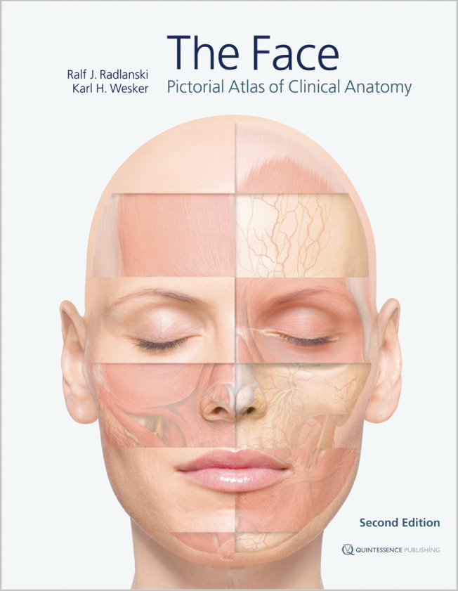
Kaufen bei QP Deutschland Kaufen bei QP United Kingdom Kaufen bei QP USAFor the first time, the highly complex topographic-anatomical relationships of facial anatomy are depicted layer by layer using extremely detailed anatomical illustrations with a three-dimensional aspect. Important landmarks, anatomical details, and clinically relevant constellations of hard and soft tissues, as well as of nerves and blood vessels, have been detailed. Another important feature is that the point of view is maintained throughout while moving through the different layers of preparation. While the accompanying text and figure captions highlight specific issues, the images remain in the foreground. The elaborate illustrations are based mainly on live anatomy and corresponding images obtained from magnetic resonance imaging, with some support from anatomical preparations.
Contents
Chapter 1. The Face
• Introduction
• The face in anterior view
• The face in lateral view
• The head in vertical view
• The head in dorsal view
• The neck
• Facial expression
• The facial skeleton
• Sectional anatomy
• Schematic representations of pathways in the face
Chapter 2. The Orbital Region
• Surface topography of the orbital region
• Preseptal muscles and fat layers
• The orbital septum and the eyeball
• Vascular and nerve supply in the orbital region
• Vascular and nerve supply in the orbital region in relation to the muscles
• Sectional anatomy of the orbital region
Chapter 3. The Nasal and Midfacial Region
• Surface topography of the nasal region
• The nose in anterior view
• The nose in lateral view
• The nose in caudal view
• The nasal cavity
• The sinuses
Chapter 4. The Mouth
• Extraoral topography of the oral region
• Topographical anatomy of the oral region
• Vascular and nerve supply of the oral region
• The oral cavity
• Anatomy of the lips, teeth, periodontium and alveolar bone in sections
• The anterior oral vestibule
• Anatomy in the area around the mandibular ramus
• The temporomandibular joint
• Anatomy of the oral region in sections
• Pathways of odontogenic spread of infections
Chapter 5. The Ear
Chapter 6. The Skin and Aging of the Face

Prof. Dr. Dr. Ralf J. Radlanski
Deutschland, Berlin1978–1983: Studium der Zahnheilkunde und Medizin in Göttingen und Minneapolis (Minnesota, U.S.A.). 1983–1989: Wissenschaftliche Grundausbildung im Zentrum Anatomie der Universität Göttingen (Promotion) und Ausbildung zum Fachzahnarzt für Kieferorthopädie in der Abt. Kieferorthopädie des Zentrums Zahn-, Mund- und Kieferheilkunde der Universität Göttingen. 1989: Habilitation an der Medizinischen Fakultät der Universität Göttingen. 1990–1992: Oberarzt der Abt. Kieferorthopädie des Zentrums Zahn-, Mund- und Kieferheilkunde der Universität Göttingen. Seit 1992: Professor und Direktor der Abt. Orale Struktur- und Entwicklungsbiologie an der Charité Universitätsmedizin Berlin, Campus Benjamin Franklin, Freie Universität Berlin. Gastprofessor an der University of California at San Francisco und an der Universität Turku (Finnland). Seit 1992: als Kieferorthopäde teilzeit in Gemeinschaftspraxis tätig. Seit 2012: Präsident der EurAsian Association of Orthodontists (EAO). 2015–2018: 1. Vorsitzender der Arbeitsgemeinschaft für Grundlagenforschung (AfG) der Deutschen Gesellschaft für Zahn-, Mund- und Kieferheilkunde Hauptforschungsgebiete: Craniofaciale Morphogenese, Orale Struktur- und Entwicklungsbiologie, Biologische Grundlagen der praktischen Kieferorthopädie. Zahlreiche Originalpublikationen, Buchbeiträge und Lehrbücher zu Fragen der Grundlagenforschung, der strukturbiologischen Grundlagen zahnärztlichen Handelns und praktische klinische Beiträge aus dem Bereich der Kieferorthopädie. Referententätigkeit zur kieferorthopädischen Weiterbildung in verschiedenen Fortbildungsinstituten. Mitglied in verschiedenen wissenschaftlichen Fachgesellschaften und Fachredaktionen.

Karl H. Wesker
Deutschland, BerlinKarl H. Wesker ist bildender Künstler und Illustrator. Er beschäftigt sich seit mehreren Jahrzehnten mit der Darstellung und didaktischen Umsetzung komplexer anatomischer Sachverhalte. Er entwickelte neue Verfahren, die zu detailgenauen und gleichzeitig ästhetisch faszinierenden Bildern der menschlichen Anatomie führen. Diese Verfahren fanden Eingang in das von Karl Wesker maßgeblich mitgestaltete dreiteilige Anatomiewerk "Prometheus" des Thieme-Verlags. Dieses Werk wurde bereits mehrfach ausgezeichnet und in zahlreichen Ländern lizenziert.


