Implantologie, 2/2025
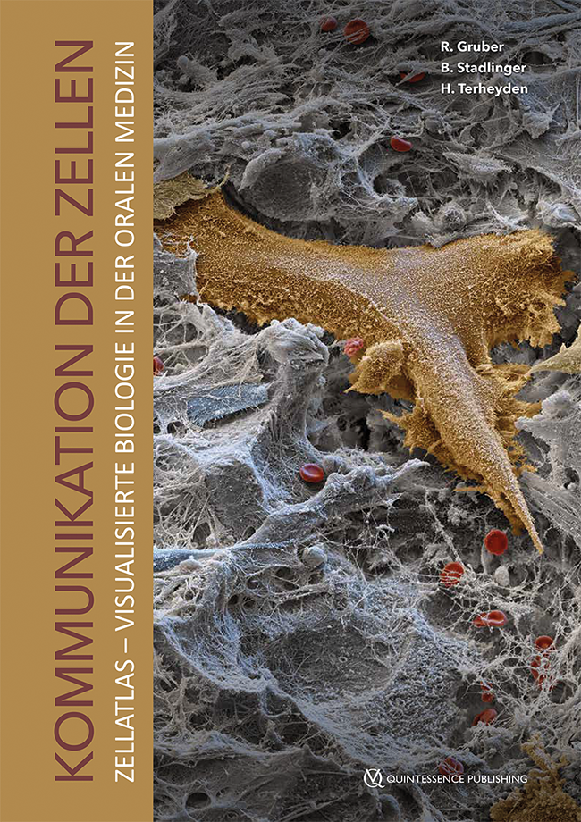
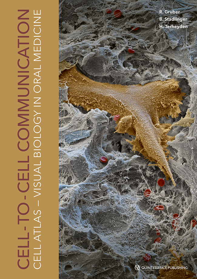
Pages 125-134, Language: GermanGruber, ReinhardDie dentale Implantologie hat sich als bewährte Methode der oralen Rehabilitation erwiesen. Histologische Analysen unterstreichen die sequenziellen Prozesse, die vom Blutgerinnsel über das Granulationsgewebe bis hin zum Geflecht- und Lamellenknochen ablaufen. Die direkte Bindung des neu gebildeten Knochens an die Implantatoberfläche, die Osseointegration, ist letztlich der Schlüssel zum Erfolg dieser Behandlungsoption. Die histologische Darstellung der Osseointegration liefert jedoch nicht nur Antworten, sie wirft auch Fragen zu den zellulären und molekularen Mechanismen der Knochenregeneration auf. Im folgenden Artikel werden diese Mechanismen mit besonderem Schwerpunkt auf der Osseointegration – am Beispiel der durch Osteoklasten verursachten katabolen Phase und der durch Osteoblasten herbeigeführten anabolen Phase – dargestellt. Besonders hervorzuheben sind hier die Osteozyten und ihre Bedeutung in der Kommunikation mit den ausführenden Zellen, den Osteoklasten und Osteoblasten. Da die zelluläre Kommunikation über das Senden und Empfangen von Signalmolekülen erfolgt, werden die Faktoren „Receptor activator of nuclear factor-kappa B ligand“ (RANKL) und Sklerostin als Beispiele erwähnt und ihre biologische und klinische Bedeutung erläutert. Die Umsetzung der biologischen Prinzipien der zellulären Kommunikation in klinische Entscheidungen in der Implantologie hat bereits begonnen.
Keywords: Implantologie, Osseointegration, Regeneration, Histologie, Osteoblasten, Osteoklasten, Osteozyten, RANKL, Sklerostin
The International Journal of Oral & Maxillofacial Implants, 2/2023
DOI: 10.11607/jomi.9709, PubMed ID (PMID): 37083911Pages 226-238, Language: EnglishMagrin, Gabriel Leonardo / Scarduelli, Letícia Daros / Rivero, Elena Riet Correa / Magini, Ricardo de Souza / Gruber, Reinhard / Benfatti, Cesar Augusto MagalhãesPurpose: To compare different socket sealing approaches for alveolar ridge preservation and assess the dimensional changes and histologic characteristics of soft and hard tissues in a 4- to 6-month period.
Material and Methods: A total of 22 patients with indicated single-tooth extraction in the maxillary nonmolar region were eligible for this study. After CBCT scanning and minimally traumatic tooth extraction, the alveolar sockets were filled with demineralized bovine bone mineral with collagen (DBBM-C) in patients from all groups except for those in the control group. Patients were divided into groups for socket sealing as follows: unsealed/spontaneous healing (control; n = 6), collagen matrix (n = 5), collagen membrane (n = 5), and autogenous graft (n = 6). A second CBCT scan was taken 4 to 6 months after extraction, and a trephine biopsy of soft and hard tissues was collected during implant placement. Tomographic dimensional changes were compared between groups. Intragroup tomographic evaluation and histological analysis were also performed.
Results: Analysis of dimensional changes did not detect differences between the socket sealing groups (P > .05). In an intragroup evaluation, the height of the buccal bone and cross-sectional area of the alveolar ridge were significantly lower 4 to 6 months after extraction for the control group (P = .031). Histological analysis revealed that the socket sealing approach had no impact on hard and soft tissue formation.
Conclusion: The data from the present study suggest that socket sealing with a collagen matrix, a collagen membrane exposed to the oral cavity, or an autogenous punch graft had no difference in the effects on volumetric maintenance and tissue formation in a period of 4 to 6 months.
Keywords: alveolar process, alveolar ridge preservation, biomaterials, histology, periodontology, randomized clinical trial, socket sealing, tomographic analysis, tooth extraction, tooth socket
International Journal of Oral Implantology, 3/2021
PubMed ID (PMID): 34415129Pages 285-302, Language: EnglishFujioka-Kobayashi, Masako / Miron, Richard J / Moraschini, Vittorio / Zhang, Yufeng / Gruber, Reinhard / Wang, Hom-LayPurpose: To investigate the effect of platelet-rich fibrin on bone formation by investigating its use in guided bone regeneration, sinus elevation and implant therapy.
Materials and methods: This systematic review and meta-analysis were conducted and reported in accordance with the Preferred Reporting Items for Systematic Reviews and Meta-Analyses guidelines. The eligibility criteria comprised human controlled clinical trials comparing the clinical outcomes of platelet-rich fibrin with those of other treatment modalities. The outcomes measured included percentage of new bone formation, percentage of residual bone graft, implant survival rate, change in bone dimension (horizontal and vertical), and implant stability quotient values.
Results: From 320 articles identified, 18 studies were included. Owing to the heterogeneity of the investigated parameters, a meta-analysis was only possible for sinus elevation. There is a general lack of data from comparative randomised clinical trials evaluating platelet-rich fibrin for guided bone regeneration procedures (only two studies), with no quantifiable advantages in terms of new bone formation or dimensional bone gain found in the platelet-rich fibrin group. For sinus elevation, the meta-analysis demonstrated no advantage in terms of histological new bone formation in the control group (bone graft alone) compared with the test group (bone graft and platelet-rich fibrin). Two studies demonstrated that platelet-rich fibrin may shorten healing periods prior to implant placement. Platelet-rich fibrin was also shown to slightly enhance primary implant stability (implant stability quotient value < 5) as assessed using implant stability quotients and resonance frequency analysis parameters, with no histological data evaluating bone–implant contact yet available on this topic. In one study, platelet-rich fibrin was shown to improve the clinical parameters when utilised as an adjunct for the treatment of peri-implantitis.
Conclusions: In the majority of studies, platelet-rich fibrin offered little or no clear advantage in terms of new bone formation as evaluated in various studies on guided bone regeneration and sinus elevation, nor in implant stability and treatment of peri-implantitis. Various authors and systematic reviews on the topic have now expressed criticism of the various study designs and protocols, and the lack of appropriate controls and available information regarding patient selection. Well-controlled human studies on these specific topics are required.
Conflict-of-interest statement: Richard J Miron holds intellectual property on platelet-rich fibrin.
All other authors declare no conflicts of interest.
Keywords: biomaterials, bone graft, growth factors, platelet-rich fibrin, platelet concentrates
International Journal of Oral Implantology, 2/2021
PubMed ID (PMID): 34006080Pages 181-194, Language: EnglishMiron, Richard J / Fujioka-Kobayashi, Masako / Moraschini, Vittorio / Zhang, Yufeng / Gruber, Reinhard / Wang, Hom-LayPurpose: To investigate the use of platelet-rich fibrin for alveolar ridge preservation compared to natural healing, bone graft material and platelet-rich fibrin in combination with bone graft material.
Materials and methods: The present systematic review was conducted and reported according to the Preferred Reporting Items for Systematic Reviews and Meta-analysis guidelines. The review examined randomised controlled trials comparing the clinical outcomes of platelet-rich fibrin with those of other modalities for alveolar ridge preservation. Studies of third molar extraction site healing were excluded. The studies were classified into three categories: natural wound healing vs platelet-rich fibrin; bone graft material vs platelet-rich fibrin; and bone graft material vs bone graft material and platelet-rich fibrin.
Results: From 179 articles identified, 16 randomised controlled trials were included. Owing to the heterogeneity of the investigated parameters, it was not possible to perform a meta-analysis. In total, 10 randomised controlled trials compared platelet-rich fibrin to natural wound healing, with seven of these demonstrating favourable outcomes to either limit postextraction dimensional changes or improve new bone formation in the platelet-rich fibrin group. Three of four studies comparing healing with bone graft material to platelet-rich fibrin found that the latter led to significantly greater horizontal or vertical bone resorption, and the bone graft material was more able to maintain the ridge dimensions. Two out of three randomised controlled trials investigating healing with both bone graft material and platelet-rich fibrin reported better outcomes using this combined approach than with bone graft material alone. All studies investigating soft tissue healing with platelet-rich fibrin demonstrated better outcomes in the platelet-rich fibrin group.
Conclusions: The majority of studies comparing healing with platelet-rich fibrin to natural healing concluded that the former more successfully limits postextraction dimensional changes than the latter. However, 75% of studies investigating platelet-rich fibrin vs bone graft material reported better results in the bone graft group with respect to its ability to maintain postextraction dimensional changes. The addition of platelet-rich fibrin to bone graft material may improve clinical outcomes, although data are limited.
Keywords: advanced platelet-rich fibrin, alveolar ridge preservation, biomaterials, extraction site management, growth factors, leucocyte- and platelet-rich fibrin, platelet concentrates, platelet-rich fibrin, systematic review
Conflict-of-interest statement: Richard J Miron holds intellectual property on platelet-rich fibrin. All other authors declare no conflicts of interest related to this study.
The International Journal of Oral & Maxillofacial Implants, 6/2020
Pages 1218-1228, Language: EnglishSadilina, Sofya / Kalakutskiy, Nikolay / Petropavlovskaya, Olga / Rumakin, Vasiliy / Gruber, ReinhardPurpose: The purpose of this prospective clinical study was to evaluate the efficiency of alveolar ridge reconstruction with the lateral border of scapula (LBS) prior to implant placement and to assess onlay graft retention and bone resorption during a short term of function.
Materials and Methods: A total of 25 partially or fully edentulous patients with severe alveolar bone atrophy received ridge reconstruction with grafts harvested from the LBS. Histologic analysis of bone grafts was performed. Six months after augmentation, patients underwent CBCT and received dental implants. After another 3 months, healing abutments and implant-supported dentures were placed. Patients were followed for an average of 24 months.
Results: Thirteen patients received primary bone grafting from LBS. Twelve patients experienced unsuccessful ridge reconstruction with other grafts before and were secondarily augmented with LBS. The average dimensions of LBS grafts were 6.3 × 2.3 × 1.2 cm. Histologic analysis confirmed the cortical nature of the graft. No donor-site complications occurred, and arm movements were restored within 2 weeks. Following augmentation, two patients had sutures disrupted that healed uneventfully after revision. The average resorption of LBS grafts after 6 months was 12.2% ± 3.0%. At the time of implant placement, the dimension of the ridge was 12.3 ± 2.0 mm and 6.9 ± 1.6 mm in height and width, respectively. The survival rate of the 174 implants placed was 98.3%.
Conclusion: LBS can be used as an alternative extraoral grafting site for extensive ridge reconstruction prior to implant placement.
Keywords: autologous bone grafts, lateral border of scapula, ridge reconstruction, severe bone atrophy
The International Journal of Oral & Maxillofacial Implants, 5/2020
DOI: 10.11607/jomi.8082, PubMed ID (PMID): 32991641Pages 917-923, Language: EnglishViteri-Agustín, Iratxe / Brizuela-Velasco, Aritza / Lou-Bonafonte, Jose Manuel / Jiménez-Garrudo, Antonio / Chávarri-Prado, David / Pérez-Pevida, Esteban / Benito-Garzón, Lorena / Gruber, ReinhardPurpose: Compaction of particulated grafts is done manually; thus, the effect of compression force on bone regeneration remains unclear. The aim of this study was to evaluate the impact of two different compression forces on the consolidation of particulated bovine hydroxyapatite.
Materials and Methods: Two titanium cylinders were fixed on the calvarium of eight New Zealand rabbits. Both defects were filled with particulated bovine hydroxyapatite subjected to a compression force of 0.7 kg/cm2 or 1.6 kg/cm2 before being covered with a resorbable collagen membrane. A handheld device that uses a spring to control the compression force applied by the plugger was used. At 6 weeks, histomorphometry of the area immediately adjacent to the calvaria bone and to the collagen membrane was performed.
Results: It was shown that next to the calvaria, the bone volume per tissue volume (BV/TV) was 29.0% ± 8.8% and 27.6% ± 8.2% at low and high compression force, respectively; the bone-to-biomaterial contact (BBC) was 58.2% ± 25.0% and 69.3% ± 22.9%, respectively (P > .05). In the corresponding area next to the collagen membrane, BV/TV was 4.9% ± 5.1% and 5.7% ± 4.7%, and the BBC was 18.3% ± 20.8% and 20.1% ± 15.9%, respectively (P > .05). In addition, the number and area of blood vessels were not significantly affected by compression force.
Conclusion: Both compression forces applied resulted in similar consolidation of bovine hydroxyapatite expressed by new bone formation and vascularization based on a rabbit calvaria augmentation model.
Keywords: bone regeneration, compaction, compression force, particulated bone graft
Quintessenz Zahnmedizin, 12/2020
Orale MedizinPages 1420-1427, Language: GermanGruber, ReinhardDie inflammatorische Osteolyse beschreibt jenen Prozess, bei dem eine chronische Entzündung
die Entstehung von knochenresorbierenden Osteoklasten begünstigt und damit letztlich zur Zerstörung des Hartgewebes führt. Wir erörtern hier, wie die chronische Entzündung mit der Osteoklastogenese auf molekularer und zellulärer Ebene gekoppelt ist. Im Prinzip fördern die Entzündungsfaktoren die vermehrte Expression von RANKL – dem Schlüsselmolekül der Osteoklastogenese – in mesenchymalen Zellen. Bei den mesenchymalen Zellen stehen die Osteozyten als Produzenten von RANKL im wissenschaftlichen Fokus. Zudem können auch Subpopulationen der T-Zellen durch deren Expression von RANKL die Osteoklastogenese
fördern. Angesprochen wird außerdem die durch die therapeutische Neutralisierung von RANKL bewirkte Verschiebung der Stammzellen zu inflammatorischen Makrophagen im Zusammenhang mit der Osteonekrose sowie die Bedeutung der Makrophagen bei der Auflösung von Entzündungen und bei der Knochenregeneration.
Keywords: Osteoklasten, Immunologie, Osteolyse, RANKL, präklinische Modelle
Quintessenz Zahnmedizin, 12/2020
Zahnmedizin allgemeinPages 1430-1435, Language: GermanTerheyden, Hendrik / Stadlinger, Bernd / Gruber, ReinhardOsteoklasten wurden lange für mesenchymale Knochenzellen gehalten. Aber neue Kenntnisse aus der Entwicklungsbiologie hinsichtlich der molekularen Steuerung der Zelldifferenzierung und der Immunmodulation des Knochenabbaus weisen die Osteoklasten klar als Mitglieder des Immunsystems aus. Osteoklasten sind für viele zahnärztliche Therapien wie die geordnete Zahneruption und kiefer-orthopädische Zahnbewegung, den Umbau eines Knochentransplantates oder die Osseointegration eines Zahnimplantates unerlässlich. Die Bedeutung der Osteoklasten bei der oralen Immunabwehr wird insbesondere bei Dysfunktionen der Osteoklasten deutlich, so beispielsweise bei der medikamenten-assoziierten Kiefernekrose. Die Immunmodulation der Osteoklastogenese kann zu foudroyanten Verläufen bis hin zur eitrigen Entzündung führen, wofür die entzündliche Wurzelresorption und die Periimplantitis klinische Beispiele sind. Die Aufgabe des Zahnarztes in diesen Situationen ist die Minderung der Osteoklastogenese durch Reduktion der mikrobiellen Antigenbelastung durch endodontische Behandlung bzw. Kontrolle des Biofilmes. Die marginale Parodontitis kann der Osteoimmunologie als Modell zur Erforschung der entzündlichen Knochenresorptionen dienen. Hiervon profitieren im Gegenzug auch andere Fachbereiche wie z. B. die Rheumatologie.
Keywords: Osteoklasten, Zelldifferenzierung, Osteoimmunologie, Wurzelresorption, Kiefernekrose, Zytokine
International Poster Journal of Dentistry and Oral Medicine, 5/2019
SupplementPoster 2102, Language: EnglishMagrin, Gabriel Leonardo / Freitas-Rafael, Stela Neuza / Passoni, Bernardo Born / Gruber, Reinhard / Benfatti, Cesar Augusto Magalhães / Peruzzo, Daiane CristinaGuided virtual surgery (GVS) has as premise a better accuracy for dental implants placement. However, the reproducibility of the implant planned position by means of surgical guides is still under investigation. This study had as objective to assess the angular and the linear (point of entry and apical extremity) deviations of single-tooth dental implants placed by two different techniques: GVS with stereolithographic guide and conventional surgery (CS) with handmade guide. This split-mouth randomized clinical trial was approved by the Research Ethical Committee involving Human Beings of the Federal University of Santa Catarina (CEPSH - UFSC; Protocol no. 1,658,040/2016) and was registered on the Brazilian Register of Clinical Trials (register number: RBR-2h745w). Twelve patients with bilateral homologous single-tooth missing in posterior mandible were selected (n=24). After scan appliance manufacturing and cone beam computed-tomography (CBCT) by dual scanning technique, cases were virtually planned and forwarded to a prototyping center to stereolitographic surgical guide manufacturing (CAD/CAM). The conventional surgical guides were obtained by scan appliances' preparation. During the surgical procedure, patients were randomized on GVS or CS by a coin toss. Each side of the mandible received one implant by a different technique. After one week, patients underwent a new CBCT scan and the images were superimposed to evaluate the differences of implants positioning between planned and performed. Student's t-test was applied at a significance level of 5% (p=0.05). Angular deviations of GVS implants showed 2.2±1.1 degrees of discrepancy while those placed by CS presented a discrepancy of 3.5±1.6 degrees, and this difference was statistically significant (p-value=0.032). For the variables coronal and apical distances (deviations), however, the null hypothesis was accepted (p-value>0.05), that is, the means between the groups of GVS and CS were not different statistically. It can be concluded that single-tooth implant placement by GVS is more accurate, at least for the angular deviation, when compared to CS with a surgical guide made by hand. Considering the linear deviations (cervical extremity and apical end), the difference between both groups cannot be demonstrated in this study. More randomized clinical trials with larger samples are needed to confirm such trends and to provide solid evidence on the subject.
Keywords: dental implants, computer-assisted surgery, oral surgery, cone-beam computed tomography, single-tooth dental implant
The International Journal of Oral & Maxillofacial Implants, 5/2018
DOI: 10.11607/jomi.6478, PubMed ID (PMID): 30231105Pages 1149-1154, Language: EnglishBruckmoser, Emanuel / Gruber, Reinhard / Steinmassl, Otto / Eder, Klaus / Watzinger, Franz / Bayerle-Eder, Michaela / Jesch, PhilipPurpose: To evaluate the sinus membrane perforation and implant survival rate after crestal minimally invasive sinus floor augmentation using hydraulic pressure and vibrations.
Materials and Methods: In this retrospective single cohort study, all patients who underwent minimally invasive sinus floor augmentation between 2007 and 2015 using hydraulic pressure and vibrations were included. The sinus membrane is elevated by physiologic saline at 1.5 bar. The fluid is then set into vibration to further separate the sinus membrane from the bony floor. The endpoints were sinus membrane perforation and the survival rate of implants.
Results: The hydraulic pressure and vibration technique was applied in 156 patients. Seven patients with perforations of the sinus membrane were treated with the lateral window approach and excluded from the follow-up analysis. In the remaining 149 patients, 184 crestal sinus floor augmentations were performed and 184 implants were placed. In 10 of these 184 cases, a perforation was suspected in the postoperative computed tomography (CT) scan. In total, the perforation rate was 8.9% (17/191). Nineteen implants were lost during the follow-up period ranging from 0.2 to 8.4 years with a median of 2.3 years. The cumulative implant survival rates after 1, 3, and 5 years were 94.4%, 87.7%, and 87.7%, respectively. No severe perioperative complications were noted.
Conclusion: The hydraulic pressure and vibration technique allows a minimally invasive crestal sinus augmentation with a perforation rate less than 10% and implant survival rates of approximately 90%.
Keywords: crestal, flapless, minimally invasive, sinus augmentation, sinus elevation, transalveolar


