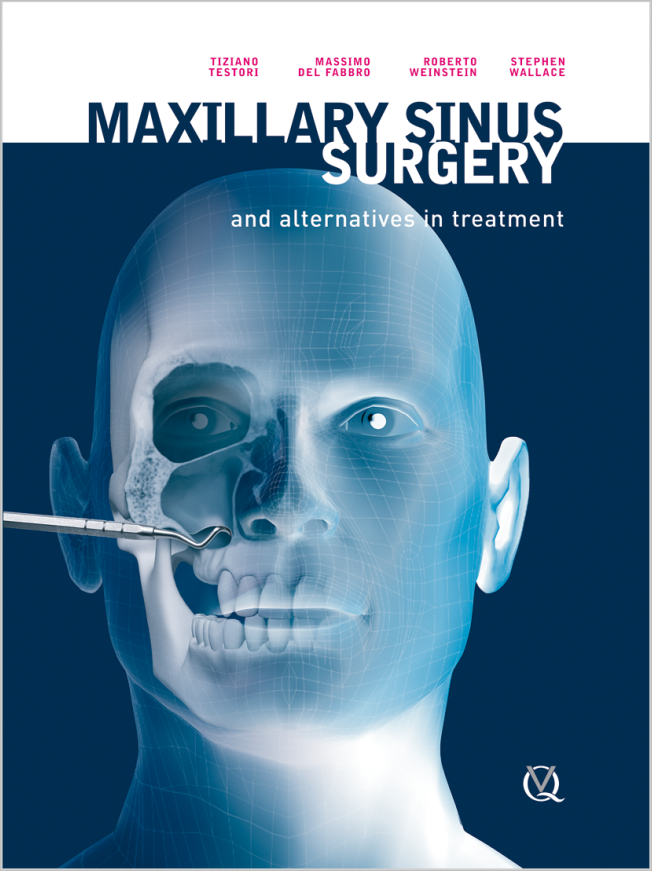
The International Journal of Oral & Maxillofacial Implants, Pre-Print
DOI: 10.11607/jomi.11434Juni 24, 2025,Seiten: 1-24, Sprache: EnglischMenini, Maria / Canullo, Luigi / Pesce, Paolo / Bixio, Francesca / Bagnasco, Francesco / Del Fabbro, MassimoBackground: Implant rehabilitation has become one of the primary treatment options for both partial and total edentulism in contemporary dentistry. In the posterior maxilla, a challeng frequently arises from insufficient available bone volume; to overcome this limitation, sinus floor elevation (SFE) procedures are often necessary. Two main techniques are employed for this purpose: the lateral window approach and the transcrestal one. Purpose: to evaluate patient-reported outcome measures related to maxillary sinus augmentation and alternative options for the rehabilitation of atrophic posterior maxilla. Study Selection: An electronic search was conducted on PubMed, Scopus and Cochrane CENTRAL up to January 31st, 2025 to identify randomized and non-randomized comparative clinical studies involving at least one group treated with maxillary SFE, with a minimum of 5 patients per group, in which PROMs were assessed. The focused question was: In patients requiring rehabilitation of atrophic posterior maxilla, is there a protocol superior to others among SFE using lateral or transcrestal approach with or without grafting, and implant placement without augmentation, in terms of patient-reported outcome measures? Pairwise meta-analysis was undertaken to estimate the overall effect when at least two studies with similar treatment comparisons, reporting the same outcome, were found. The primary outcome was postoperative pain, measured using the VAS scale in the first seven days after surgery. Secondary outcomes were any other PROMs like postoperative symptoms or standard satisfaction questionnaires. Results: The electronic search initially identified 104 articles. After deduplication, 56 articles were screened, and 12 studies (12 RCTs and 1 non-RCT) were finally included. Ten studies were judged at moderate risk of bias and one RCT at high risk. The non-RCT study was judged at moderate risk of bias. Meta-analysis could be performed only for the proportion of patients reporting pain at 1, 3 and 7 days postoperatively. The graftless approach resulted in less pain at 3- and 7-days post-op compared to SFE with graft. Transcrestal SFE also showed less post-op discomfort than lateral SFE. Conclusions: The results indicated that patients may experience reduced discomfort and inflammation when graftless procedures and transcrestal approaches are used. Overall, there is a paucity of evidence regarding the assessment of PROMs in the rehabilitation of atrophic posterior maxilla.
Schlagwörter: Bone graft, Bone substitute, Sinus lift
The International Journal of Oral & Maxillofacial Implants, 6/2024
DOI: 10.11607/jomi.10803, PubMed-ID: 38394440Seiten: 851-856, Sprache: EnglischOhayon, Laurent / Del Fabbro, MassimoPurpose: To present a sling suture technique used to stabilize a collagen membrane against the lateral bone window to improve bone substitute stability inside the sinus cavity. Materials and Methods: Maxillary sinus floor augmentation was performed on 17 patients (8 women and 9 men; mean age 58.2 years) using a lateral approach with the sling suture technique to maintain a collagen membrane against the lateral bone window. Postoperative CBCT images were captured at 6-month follow-up of each patient to monitor the bone graft stability at the level of the lateral antrostomy. Clinical postoperative pain and swelling were assessed via visual analog scale (VAS) questionnaire, measured from level 1 (low) to level 5 (acceptable) to level 10 (high) at 1 week postoperative. Results: No bone substitute displacement was observed in any clinical cases on the CBCT images at 6 months postoperative. The pain and swelling levels observed 1 week postoperatively were significantly low (mean ± SD; 1.6 ± 1.0 and 2.1 ± 0.9, respectively). Conclusions: The use of the sling suture technique to maintain a barrier membrane at the level of the lateral bone window in cases of maxillary sinus floor augmentation using a lateral approach is a predictable protocol to prevent bone substitute displacement outside the sinus cavity.
Schlagwörter: bone substitute, case series, CBCT collagen membrane, sinus floor elevation, suture
International Journal of Periodontics & Restorative Dentistry, 6/2024
DOI: 10.11607/prd.6778, PubMed-ID: 38198431Seiten: 663-672, Sprache: EnglischPradella, Sandro / Morellini, Chiara / Formentini, Damiano / del Fabbro, MassimoA total of 57 interproximal restorations invading the supracrestal tissue attachment were evaluated in terms of crestal bone loss over a mean period of 15 years (10 to 23 years). The distance from the cavity margin to the bone was measured at T0 (after the restoration; baseline) and controlled using radiographs and a measurable landmark. The mean vertical bone loss was 0.46 mm, with a 96.49% survival rate. Smoking habits (P = .02) and tooth type (P = .03) significantly affected bone loss. The proposed technique could help the clinician in adopting a minimally invasive approach in the treatment of heavily compromised teeth. Future research with rigorous study designs would be interesting to guide the clinical decision-making.
Schlagwörter: bone loss, crown lengthening, deep margins, direct restoration, prosthetic dentistry, restorative dentistry
The International Journal of Oral & Maxillofacial Implants, 5/2024
DOI: 10.11607/jomi.10723, PubMed-ID: 38607361Seiten: 674-683, Sprache: EnglischGuarnieri, Renzo / Testarelli, Luca / Galindo-Moreno, Pablo / Del Fabbro, Massimo / Testori, TizianoPurpose: To describe the emerging evidence concerning etiologic factors and pathophysiologic mechanisms involved in peri-implant inflammatory diseases. Materials and Methods: An electronic search for articles published until November 2022 was conducted in MEDLINE by three independent reviewers to identify the manuscripts’ reporting data on etiologic factors and pathophysiologic mechanisms associated with peri-implant diseases. Results: Current evidence suggests that peri-implant mucositis and peri-implantitis are inflammatory conditions linked to a microbial challenge. However, in recent years there has been increasing evidence indicating that certain peri-implant inflammatory conditions may not be primarily related to biofilm-mediated infectious processes but rather to other biologic mechanisms, such as a foreign body response. Conclusions: Current evidence in the dental and medical literature opens new avenues for a more complex interpretation of the etiopathogenetic factors involved in peri-implant diseases. A better understanding of various factors related to the host response, including dysbiosis mechanisms associated with changes in microbiota composition, is necessary for a more precise physiopathologic characterization of these diseases.
Schlagwörter: dental implant, foreign body response, microbial biofilm, peri-implant diseases, peri-implant infection
International Journal of Oral Implantology, 2/2024
PubMed-ID: 38801332Seiten: 189-198, Sprache: EnglischTestori, Tiziano / Scaini, Riccardo / Friedland, Bernard / Saibene, Alberto Maria / Felisati, Giovanni / Craig, John R / Deflorian, Matteo / Zuffetti, Francesco / Del Fabbro, Massimo / Wang, Hom-LayMaxillary sinus grafting is a predictable regenerative technique to facilitate maxillary posterior implant placement when there is insufficient vertical bone height inferior to the maxillary sinuses to allow placement of implants of adequate dimensions. It enables an increase in vertical bone height, which makes implant placement easier. Maxillary sinus mucosal membrane perforation is one of the most common intraoperative complications during maxillary sinus grafting and may result in extrusion of graft material into the sinus. When this occurs, the mucociliary function of the maxillary sinus may expel the extruded graft material through its natural ostium, though graft particles may remain in the sinus or possibly occlude the natural ostium. After grafting, transient maxillary sinus mucosal oedema may occur. A postoperative CBCT scan may reveal varying degrees of sinus opacification, namely partial, subtotal or total. Although it is always possible to identify graft material, which may enter the sinus as a result of membrane perforation that might not even be visible to the implantologist during the surgical procedure, it is challenging to assess whether sinus opacification is due to mucosal thickening or mucus accumulation. The aim of the present case series was to offer a pragmatic approach to managing asymptomatic patients whose CBCT scans demonstrated partial, subtotal or total maxillary sinus opacification with bone graft particles that seemed to have been extruded into the sinus.
Schlagwörter: maxillary antrum, maxillary sinus elevation, maxillary sinus grafting, osteomeatal complex, osteomeatal unit, sinus membrane perforation, sinus mucosa, sinus opacification, transient swelling
The authors declare no conflicts of interest relating to this study.
International Journal of Esthetic Dentistry (DE), 2/2024
Clinical ResearchSeiten: 124-136, Sprache: DeutschGalli, Fabio / Deflorian, Matteo / Zucchelli, Giovanni / Del Fabbro, Massimo / Testori, TizianoEine retrospektive Studie mit 5 bis 13 Jahren NachbeobachtungZiel: In der vorliegenden retrospektiven Studie sollten die klinischen Ergebnisse von festsitzendem, auf den Prinzipien der biologisch orientierten Präparationstechnik (BOPT) basierenden Zahnersatz über mittlere bis lange Beobachtungszeiträume ausgewertet werden.
Material und Methoden: Die Behandlungsdokumentation von Patienten, die zwischen Januar 2007 und Dezember 2014 festsitzend rehabilitiert worden waren, wurden retrospektiv ausgewertet. Patienten, deren Akten die Einschlusskriterien erfüllten, wurden außerdem zu einer abschließenden Kontrolluntersuchung einbestellt. Ausgewertet wurden die parodontalen Parameter, das Vorliegen von Gingivarezessionen sowie die aufgetretenen technischen und biologischen Komplikationen.
Ergebnisse: Insgesamt 58 Patienten wurden per Recall eingeladen. Von diesen konnten 52 Patienten mit 220 überkronten Zähnen ausgewertet werden (Ausfallrate: 13,8 %). Die durchschnittliche Beobachtungsdauer waren 9,3 Jahre (Spannweite 5 bis 13 Jahre): 14 Patienten (114 überkronte Zähne) waren 5 bis 8 Jahre, 36 Patienten (106 überkronte Zähne) 9 bis 13 Jahre nachbeobachtet worden. Die prothetische Überlebensrate betrug 99,6 %. An Komplikationen waren eine Wurzelfraktur (0,4 %) und vier Verblendkeramikabplatzungen (1,8 %) dokumentiert. Sechs Zähne (2,7 % der untersuchten Kronen) zeigten eine Gingivarezession von < 1 mm und 13 Zähne (24 Stellen) wiesen eine Sondierungstiefe von 4 mm auf (5,9 % der prothetischen Kronen/1,8 % der sondierten Stellen). Schließlich wurde an 20 Stellen (1,5 %) bzw. 7 Zähnen (3,2 %) Sondierungsbluten beobachtet.
Schlussfolgerungen: Die Präparation nach den BOPT-Regeln (d. h. mit vertikaler, kantenfreier Präparation) führte mittel- bis langfristig zu einem gesunden Parodont und stabiler Gingiva. Das prothetische Behandlungsergebnis blieb dauerhaft erhalten.
Schlagwörter: biologisch orientierte Präparationstechnik, BOPT, parodontale Gesundheit, Gingivarezession, Weichgewebestabilität, retrospektive Studie
International Journal of Esthetic Dentistry (EN), 2/2024
Clinical ResearchPubMed-ID: 38726854Seiten: 112-124, Sprache: EnglischGalli, Fabio / Deflorian, Matteo / Zucchelli, Giovanni / Del Fabbro, Massimo / Testori, TizianoA 5- to 13-year follow-up retrospective studyAim: The present study was a retrospective medium- to long-term follow-up assessment of the clinical outcomes of patients rehabilitated with fixed prostheses according to the biologically oriented preparation technique (BOPT) principles.
Materials and methods: Clinical records of patients rehabilitated between January 2007 and December 2014 were retrospectively assessed. Patients whose records met the inclusion criteria were also recalled for a hygiene visit. Data analyzed included the patients’ periodontal condition, the presence of gingival recessions as well as any technical or biologic prosthetic complication.
Results: Fifty-eight patients were recalled; of these, 52 patients who had received 220 crowns were available for the evaluation (the dropout being 13.8%). The average follow-up was 9.3 years (range 5 to 13 years): 14 patients (114 prosthetic crowns) had a follow-up between 5 and 8 years, and 36 patients (106 prosthetic crowns) between 9 and 13 years. The prosthetic survival rate was 99.6%. One radicular fracture (0.4%) and four chippings of the veneering porcelain (1.8%) were recorded. Six teeth (2.7% of the examined prosthetic crowns) presented gingival recession of < 1 mm, and 13 teeth (24 sites) had a pocket probing depth of 4 mm (5.9% of the prosthetic crowns/1.8% of the sites). Finally, 20 sites (1.5%) in seven teeth (3.2%) showed bleeding on probing.
Conclusions: Tooth preparation according to the BOPT principles (ie, with a vertical finishing line) resulted in medium- to long-term periodontal health and stability of the gingival tissue, and prosthetic success was maintained.
Schlagwörter: BOPT, periodontal health, gingival recession, soft tissue stability, retrospective study
International Journal of Periodontics & Restorative Dentistry, 2/2023
DOI: 10.11607/prd.6463, PubMed-ID: 37232685Seiten: 241-246, Sprache: EnglischOhayon, Laurent / Del Fabbro, MassimoThe aim of this retrospective analysis was to evaluate the clinical and radiographic results of a shortened protocol (using a lateral approach) for early surgical reentry, following a large sinus membrane perforation that occurred during maxillary sinus augmentation (lateral approach), for the rehabilitation of patients with an atrophic posterior maxilla. Between May 2015 and October 2020, seven patients underwent reentry surgery using a lateral approach protocol 1 month after a large sinus membrane perforation during maxillary sinus floor augmentation with lateral approach surgery. All patients presented a residual under-sinus bone height < 3 mm in the posterior maxilla. The sinus membrane was elevated during the reentry surgery, without any difficulty for any patient, using manual blunt elevators or piezoelectric devices, and the sinus floor height was augmented with bone substitute particles. No further perforations were made, and no complications were recorded during the follow-up period from 18 months to 6 years. The 1-month waiting period after the initial sinus surgery allows easy sinus membrane elevation and a lack of complications. This timing could be a feasible option for surgical reentry after a large sinus membrane perforation occurs.
The International Journal of Oral & Maxillofacial Implants, 5/2022
DOI: 10.11607/jomi.9710Seiten: 1003-1025, Sprache: EnglischDel Fabbro, Massimo / Pozzi, Alessandro / Romeo, Davide / de Araújo Nobre, Miguel / Agliardi, EnricoPurpose: To evaluate the performance of fixed complete dental prostheses supported by axial and tilted implants after at least 3 years of follow-up.
Materials and Methods: An electronic search plus a hand search up to April 2021 was undertaken. Clinical studies were selected using specific inclusion criteria, independent of the study design. The main outcomes were cumulative implant survival rate, marginal bone level changes, and complications, after ≥ 3 years of follow-up. The difference in outcomes between axial and tilted implants and between the maxilla and mandible was evaluated using meta-analysis and the Mantel-Cox test.
Results: Out of 824 articles retrieved, 24 were included. In total, 2,637 patients were rehabilitated with 2,735 full prostheses (1,464 maxillary, 1,271 mandibular), supported by 5,594 and 5,611 tilted and axial implants, respectively. In a range between 3 and 18 years of follow-up, 274 implants failed. The cumulative implant survival rate was 93.91% and 99.31% for implants and prostheses, respectively. The mean marginal bone level change was moderate, exceeding 2 mm in only two studies. Marginal bone loss was significantly lower around axial compared with tilted implants (P < .0001), whereas it was not affected by arch (maxilla vs mandible; P = .17).
Conclusion: Fixed complete dental prostheses supported by tilted and axially placed implants represent a predictable option for the rehabilitation of edentulous arches. Further randomized trials are needed to determine the efficacy of this surgical approach and the remodeling pattern of marginal bone in the long term.
Schlagwörter: axial implants, immediate loading, mandible, marginal bone loss, maxilla, tilted implants
International Journal of Periodontics & Restorative Dentistry, 4/2022
DOI: 10.11607/prd.5833Seiten: 471-477, Sprache: EnglischTestori, Tiziano / Deflorian, Matteo / Scaini, Riccardo / Wang, I-Ching / Zucchelli, Giovanni / Steigmann, Marius / Del Fabbro, Massimo / Wang, Hom-Lay / Francetti, LucaCommon challenges encountered for atrophic maxilla rehabilitation are the inadequate width and height of attached keratinized mucosa (AKM) and shallow vestibular depth. This study presents a buccally displaced palatal (BDP) flap technique to increase the tissue thickness and AKM width at the second-stage surgery and reestablish the correct fornix depth. The peri-implant pocket depths, modified Plaque Index score, modified sulcus Bleeding Index score, and soft tissue recession were evaluated 6 and 12 months after prostheses loading. A total of 52 implants were placed and analyzed, and no implant failures were found. No significant changes in peri-implant parameters were observed between 6 and 12 months, and mean recession was less than 0.2 mm after 12 months. Though this change was statistically significant, it was clinically irrelevant. The results demonstrate that adequately healthy peri-implant soft tissues and substantial dimensional stability of vestibular soft tissues at the 1-year follow-up were achieved with the BDP flap technique. The BDP flap could represent a viable option for increasing the width and the height of AKM and establishing the correct maxillary fornix depth.


