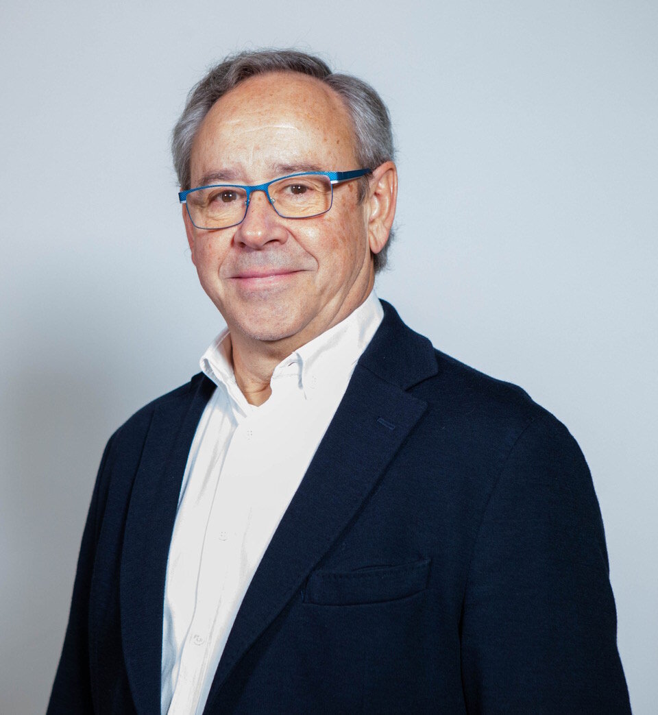
Wir verwenden Cookies ausschließlich zu dem Zweck, technisch notwendige Funktionen wie das Login oder einen Warenkorb zu ermöglichen, oder Ihre Bestätigung zu speichern. Mehr Informationen zur Datenerhebung und -verarbeitung finden Sie in unserer Datenschutzerklärung.
Mariano Sanz ist Professor und Leiter der Abteilung für Parodontologie an der Universität Complutense Madrid (Spanien) und Professor an der Fakultät für Zahnmedizin an der Universität Oslo (Norwegen). Er schloss sein Medizinstudium 1981 an der Universität Complutense Madrid ab, wo er 1983 einen Abschluss in Stomatologie erhielt und 1985 zum Doktor der Medizin promovierte. Seine Facharztausbildung in Parodontologie erhielt er 1987 an der University of California in Los Angeles (UCLA). Er wurde mit Ehrendoktorwürden von der Universität San Sebastian in Santiago (Chile), der Universität von Göteborg (Schweden) und der Universität von Coimbra (Portugal) ausgezeichnet.
Seit 2005 ist Prof. Sanz Vorsitzender der ETEP-Forschungsgruppe zur Ätiologie und Therapie parodontaler Erkrankungen, deren Hauptforschungsbereiche die orale Mikrobiologie, bakterielle Wirt-Interaktionen und antimikrobielle Ansätze in der Behandlung von Gingivitis und Parodontitis sind. Die Forschungsgruppe hat klinische Studien zur Wirksamkeit unterschiedlicher Ansätze zur parodontalen Regeneration mittels chirurgischer Protokolle unter Verwendung von Zahnimplantaten und therapeutische Ansätze zur Behandlung von Periimplantitis durchgeführt.
Prof. Sanz hat 230 Artikel in wissenschaftlichen Zeitschriften veröffentlicht, 50 Buchkapitel verfasst und war in den letzten fünf Jahren als eingeladener Referent an mehr als 200 wissenschaftlichen Veranstaltungen beteiligt. Er ist stellvertretender Redakteur des Journal of Clinical Periodontology und Evidence-Based Dental Practice und Mitglied der Redaktionsausschüsse verschiedener anderer zahnmedizinischer Zeitschriften. Er wurde mit dem Jens-Waerhaug-Forschungspreis der Skandinavischen Gesellschaft für Parodontologie (1984), dem Outstanding-Service-Preis der International Association for Dental Research (2015) und dem IADR-Straumann-Preis für regenerative parodontale Medizin (2015) ausgezeichnet.
Er ist Mitglied des Exekutivkomitees der European Federation of Periodontology (EFP) und Vorsitzender ihres Workshop-Komitees. Er hat die EFP zuvor als Präsident (1993-1994) und Generalsekretär (1998–2005) gedient. Er ist Präsident der Osteology Foundation, Präsident der kontinentaleuropäischen Abteilung der International Association for Dental Research und Präsident der Association for Dental Education in Europe (ADEE).
Länge: 17 Minuten Produktionsjahr: 2016 Sprache: Englisch, Deutsch Reihen: Cell-to-Cell Communication verfügbar seit: Februar 12, 2016
Veranstaltungen
The 15th International Symposium on Periodontics and Restorative Dentistry (ISPRD)
Juni 12, 2025 — Juni 15, 2025Boston Marriott Copley Place, Boston, MA, Vereinigte Staaten von Amerika
Referenten: Edward P. Allen, Evanthia Anadioti, Gustavo Avila-Ortiz, Christopher Barwacz, Florian Beuer, Nitzan Bichacho, Francesco Cairo, Vivianne Chappuis, Chia-Yu (Jennifer) Chen, Stephen J. Chu, Florin Cofar, Daniel Coleman, Luca Cordaro, Daniel Cullum, Luca De Stavola, Carlo Ercoli, Vincent Fehmer, Iñaki Gamborena, Maria L. Geisinger, William Giannobile, Luca Gobbato, Oscar Gonzalez-Martin, Jim Janakievski, Y. Natalie Jeong , Ronald Jung, Joseph Kan, Jihyon Kim, Gregg Kinzer, Christopher Köttgen, Ina Köttgen, Stefen Koubi, Purnima S. Kumar, France Lambert, Alejandro Lanis, Ernesto A. Lee, Paul Levi Jr, Tomas Linkevičius, Pascal Magne, Pamela Maragliano-Muniz, Konrad H. Meyenberg, Craig M. Misch, Ricardo Mitrani, Rodrigo Neiva, Giorgio Pagni, Gianluca Paniz, Giulio Rasperini, Pooyan Refahi, Andrea Ricci, Marisa Roncati, Paul S. Rosen, Irena Sailer, Mariano Sanz, Ignacio Sanz Martín, Ignacio Sanz Sànchez, Todd Scheyer, Frank Schwarz, Massimo Simion, Frank Spear, Martina Stefanini, Clint Stevens, Lorenzo Tavelli, Tiziano Testori, Sejal Thacker, Teppei Tsukiyama, Istvan Urban, Eric Van Dooren, Diego Velasquez, Hom-Lay Wang, Giovanni Zucchelli, Otto Zuhr
Quintessence Publishing Co., Inc. USA
31st EAO Annual Scientific Meeting
Details make perfectionOktober 24, 2024 — Oktober 26, 2024MiCo - Milano Convention Centre, Milano, Italien
Referenten: Bilal Al-Nawas, Gil Alcoforado, Federico Hernández Alfaro, Sofia Aroca, Wael Att, Gustavo Avila-Ortiz, Kathrin Becker, Anne Benhamou, Juan Blanco Carrión, Dieter Bosshardt, Daniel Buser, Francesco Cairo, Paolo Casentini, Raffaele Cavalcanti, Tali Chackartchi, Renato Cocconi, Luca Cordaro, Luca De Stavola, Nuno Sousa Dias, Egon Euwe, Vincent Fehmer, Alberto Fonzar, Helena Francisco, Lukas Fürhauser, German O. Gallucci, Oscar Gonzalez-Martin, Dominik Groß, Robert Haas, Alexis Ioannidis, Simon Storgård Jensen, Ronald Jung, France Lambert, Luca Landi, Georg Mailath-Pokorny jun., Silvia Masiero, Iva Milinkovic, Carlo Monaco, José Nart, José M. Navarro, Katja Nelson, Manuel Nienkemper, David Nisand, Michael Payer, Sergio Piano, Bjarni E. Pjetursson, Sven Reich, Isabella Rocchietta, Giuseppe Romeo, Irena Sailer, Mariano Sanz, Ignacio Sanz Martín, Frank Schwarz, Shakeel Shahdad, Massimo Simion, Ralf Smeets, Benedikt Spies, Bogna Stawarczyk, Martina Stefanini, Hendrik Terheyden, Tiziano Testori, Daniel Thoma, Ana Torres Moneu, Piero Venezia, Lukas Waltenberger, Hom-Lay Wang, Stefan Wolfart, Giovanni Zucchelli, Otto Zuhr
European Association for Osseintegration (EAO)
360 IMPLANTOLOGY by MIS – MIS Global Conference
September 12, 2024 — September 14, 2024Palau de Congressos de Palma, Palma de Mallorca, Spanien
Referenten: Tara Aghaloo, Serhat Aslan, Nitzan Bichacho, Darko Božić, Tali Chackartchi, Victor Clavijo, Gustavo Giordani, Moshe Goldstein, Galip Gürel, Stefen Koubi, Hilal Kuday, Alberto Monje, Stavros Pelekanos, Mario Alonso Puig, Ariel J. Raigrodski, Aušra Ramanauskaitė, Isabella Rocchietta, Mariano Sanz, Ignacio Sanz Sànchez, Venceslav Stankov
SEPA 24 Bilbao
Clinical evidence based on scientific evidenceMai 29, 2024 — Juni 1, 2024Bilbao, Spanien
Referenten: Eduardo Anitua, Sofia Aroca, Serhat Aslan, Gustavo Avila-Ortiz, Juan Blanco Carrión, Gonzalo Blasi, Nagihan Bostanci, Iain L. C. Chapple, Jan Cosyn, Glécio Vaz de Campos, Luca De Stavola, Jan Derks, Vincent Fehmer, Elena Figuero, Sergio García, Alfonso L. Gil, Oscar Gonzalez-Martin, Adrian Guerrero, Sérgio Kahn, Alejandro Lanis, Antonio Liñares, Ferrán Llansana, Francesco Mangano, Dino Calzavara mantovani, Mauro Merli, Juan Mesquida, Alberto Monje, Eduardo Montero, Stefano Parma-Benfenati, Bjarni E. Pjetursson, Pablo Ramírez, Mariano Sanz, Ignacio Sanz Sànchez, Beatriz Solano Mendoza, Jacobo Somoza, Martina Stefanini, Maurizio S. Tonetti, Leonardo Trombelli, Ion Zabalegui
Zeitschriftenbeiträge dieses Autors
International Journal of Periodontics & Restorative Dentistry, Pre-Print
Surgical crown lengthening (SCL) is a predictable method of increasing the availability of supragingival tooth structure for biological, aesthetic and/or restorative reasons. However, the literature demonstrates a lack of precision when performing this common procedure due to a number of factors. Among those factors, the use of standardised measurements and reference points as well as the limitations inherent to the classical surgical protocol will be presented. This review discusses the importance of individualising supracrestal soft tissue dimensions during SCL interventions and the use of appropriate diagnostic tools while taking into account the phenotypic characteristics of the patient in both functional and esthetic crown lengthening.
Aims: The primary aim of this in vitro study was to compare methods for generating 3D-printed replicas through virtual segmentation, utilizing artificial intelligence (AI) or manual processes, by assessing accuracy in terms of volumetric and linear discrepancies. The secondary aims were the assessment of time efficiency with both segmentation methods, and the effect of post-processing on 3D-printed replicas. Methods: Thirty teeth were scanned through Cone Beam Computed Tomography (CBCT), capturing the region of interest from human subjects. DICOM files underwent virtual segmentation through both AI and manual methods. Replicas were fabricated with a stereolithography 3D printer. After surface scanning of pre-processed replicas and extracted teeth, STL files were superimposed to compare linear and volumetric differences using the extracted teeth as the reference. Post-processed replicas were scanned to assess the effect of post-processing on linear and volumetric changes. Results: AI-driven segmentation resulted in statistically significant mean linear and volumetric differences of -0.709mm (SD 0.491, P< 0.001) and -4.70%, respectively. Manual segmentation showed no statistically significant differences in mean linear, -0.463mm (SD 0.335, P<0.001) and volumetric (-1.20%) measures. Comparing manual and AI-driven segmentations, AI-driven segmentation displayed mean linear and volumetric differences of -0.329mm (SD 0.566, p=0.003) and -2.23%, respectively. Additionally, AI segmentation reduced the mean time by 21.8 minutes. When comparing post-processed to pre-processed replicas, there was a volumetric reduction of -4.53% and a mean linear difference of -0.151mm (SD 0.564, p=0.042). Conclusion: Both segmentation methods achieved acceptable accuracy, with manual segmentation slightly more accurate but AI-driven segmentation more time-efficient. Continuous improvement in AI offers the potential for increased accuracy, efficiency, and broader application in the future.
Schlagwörter: 3d printing, artificial intelligence, autotransplant, computeraided- manufacturing, digital dentistry, Stereolithography
The management of severe mucogingival deformities surrounding malpositioned implants represents a complex issue with a high risk of aesthetic failure. The present case report describes a mucogingival and restorative approach for the treatment of severe localized peri-implant tissue deficiencies with interproximal bone loss combined with an interproximal gingival recession on the adjacent natural tooth. This consists of maintaining a non-restorable malpositioned implant, submerging it through one or multiple vertical soft tissue augmentations, according to the defect severity, and delivering a tooth supported bridge involving the adjacent natural teeth. The step-by-step surgical technique adopted for implant submergence and vertical soft tissue grafting, as well as the subsequent surgical and prosthetic interventions, are described, presenting results at 5 years from implant submergence and 3 years from prosthetic finalization.
Schlagwörter: Implants, Periodontal Surgery, Prosthetic Dentistry, Gingival Recession, Mucogingival Surgery, Dental Implant, Case Report
Consensus on a valid and comprehensive set of outcomes to capture the full benefits and harms of implant dentistry interventions is key for progressing toward better clinical guidelines and policy. In this field, research remains fragmented and uses too many different outcomes. Studies are unable to cover the full breadth necessary to properly evaluate benefits, harms, and costs. Best-practice examples inspired the Implant Dentistry Core Outcome Set and Measurements (ID-COSM) initiative that identified four core outcome domain areas (pathophysiology, implant/prosthesis lifespan, life impact, and access to care), five essential outcomes mandatory for all trials, and six outcomes mandatory in specific circumstances. This innovative multistep approach combined input from scientific evidence, patients from multiple countries, methodologists, and industry representatives. The ID-COSM consensus aspires to contribute to better adoption of relevant and valid outcomes in trials and enable their results to be combined in high-quality meta-analyses to support better-informed care and policy.
Objectives: The aim of this retrospective case series was to report the performance up to 5 years of an innovative surgical design (the apically incised coronally advanced surgical technique [AICAST]) for the regenerative treatment of one- or two-walled intrabony periodontal lesions.
Method and materials: After completion of standard step I to II periodontal therapy, nine isolated periodontal defects were treated through AICAST. The following clinical outcome measurements were collected before the surgical intervention and at the last available follow-up: probing pocket depth (PPD), recession depth (REC), and clinical attachment level (CAL). Periapical radiographs of the treated teeth were also taken at baseline and at the last available follow-up (18 months or 5 years postoperatively).
Results: A mean (± standard deviation) PPD reduction of 6.05 ± 1.76 mm (P < .01), REC reduction of 1.15 ± 1.97 mm (P = .119), and CAL gain of 7.20 ± 2.13 mm (P < .01) were attained when comparing preoperative results with the last follow-up visit. CAL gain of 6 mm or more was reached in eight out of nine treated cases (88.9%), with a residual PPD of 2 to 3 mm in all the cases. Complete radiographic fill of the intrabony component was present in all the defects, while detectable suprabony radiographic filling was identified in two cases.
Conclusion: AICAST represents an innovative surgical design for the treatment of deep intrabony defects and the eventual reduction of the associated gingival recessions. Preliminary results show good performance in terms of clinical attachment gains and maintenance of the marginal tissues.
Schlagwörter: coronally advanced flap, enamel matrix derivatives (EMD), papilla preservation flap, periodontal regeneration, periodontitis
Purpose: To investigate the subgingival microbiological profiles of patients with periodontitis, to determine their stage and grade scores and to evaluate the differences in the microbiota among different stages and grades.
Materials and Methods: Sixty-seven (n = 67) periodontitis patients were selected. Periodontitis staging and grading, following the 2018 classification system, were defined. Following a clinical examination, subgingival samples were taken from the deepest periodontal pocket of each quadrant for cultivation, identification and quantification. The prevalence, proportion and counts of nine selected periodontal pathogens were determined, and differences between periodontitis stages III and IV and grades B and C were assessed.
Results: All nine cultivable periodontal bacteria were detected, of which the most prevalent was P. intermedia (91.0%) and the least prevalent were E. corrodens (9.0%) and C. ochracea (9.0%). The frequency of detection of the two main target pathogens, A. actinomycetemcomitans and P. gingivalis, was 41.8% and 76.1%, respectively. The prevalence (grade B: 80.6%, grade C: 55.6%, p = 0.035) and total counts (grade B: 19.8 colony forming units – CFU/ml-4 (1.9–52.8); grade C: 4.0 CFU/ml-4 (0.0–26.4); p = 0.022) of F. nucleatum were statistically significantly higher in grade B than in grade C periodontitis patients, whereas the counts of P. gingivalis and A. actinomycetemcomitans were similar between grades and stages.
Conclusion: Our study suggests that relevant differences between the various grades of periodontitis exist only in the numbers of F. nucleatum. Prevalence and quantities of other cultivable species between different stages and grades of periodontitis seem to be similar.
Schlagwörter: periodontitis, stage, grade, Porphyromonas gingivalis, Aggregatibacter actinomycetemcomitans, Fusobacterium nucleatum
Purpose: No information is available on the perception of the quality of care in patients treated for periodontitis. The purpose of this article was to assess how periodontitis-affected patients perceive the quality of periodontal treatment (PT) and to measure the factors which may influence it.
Materials and Methods: 306 subjects who completed PT were invited to participate. Questionnaires and visual analogic scales (VAS) evaluating perception of quality of care, symptoms, and oral health related quality of life (OHRQoL) were handed out. Oral and periodontal indicators were collected before and after treatment. The impact of different factors on perception of quality was assessed with a regression model.
Results: Quality evaluation was high yet unrelated for both patients and clinicians (p = 0.983). Quality was negatively influenced by the number of residual oral infections (p 0.001), patient's age (p = 0.07) and presence of residual pain at completion of PT (p = 0.02). Professionalism, kindness of the staff and communication skills were the characteristics mostly appreciated. The OHRQoL was influenced by the number of residual teeth (p 0.001), increasing age of patients (p = 0.08), number of residual infections (p 0.01) and pain (p = 0.04).
Conclusions: Patients' quality perception appeared to be influenced by clinical and emotional aspects. Oral care providers should be aware of the impact of non-clinical factors in patients' appreciation of quality of treatment.
Schlagwörter: oral health related quality of life, periodontitis, quality of care
This report presents a histologic assessment of guided bone regeneration for dehiscence defects treated with bovine bone mineral or a combination of autogenous and synthetic bone. The samples were obtained from an autopsy specimen donated by a patient, which is a rare opportunity to evaluate longterm results of guided bone regeneration and osseointegration. The values for bone-to-implant contact were similar in both sites. The augmentation with bovine bone mineral demonstrated bone reconstruction after 1 year, whereas the augmentation with autogenous and synthetic bone failed to maintain the augmented volume, eventually leading to mucosal recession after 5.5 years.
Root coverage in the anterior mandible is challenging due to a thin gingiva, shallow vestibule, and/or high frenulum. This case series reports on the flattening of the root surface to create a new emergence profile conceived with a two-step surgical approach aimed at providing more space for the graft, increasing the thickness of the gingival margin, and getting extra soft tissue in the open area of the recession. A total of 10 patients with recessions affecting the mandibular incisors were treated to evaluate this two-step approach, which included odontoplasty of the root followed by a connective tissue graft. At 1 year, the mean coverage was 100% in Class II recessions, and 80.5% in Class III. The mean keratinized tissue increase was 5.80 ± 1.75 mm. This surgical approach could be proposed as an alternative when treating mandibular anterior teeth with root prominence or with a buccally tilted position.