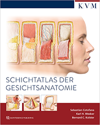
Schichtatlas der Gesichtsanatomie
Expected publication: September 2025
1st Edition 2025
Book
Hardcover; 24 x 30 cm, 302 pages, 210 illustrations
Language: German
Categories: Facial Esthetics, Plastic Surgery, Oral/Maxillofacial Surgery, ENT medicine
Stock No.: 30343
ISBN 978-3-86867-716-4
KVM Verlag
Product in preparation.
Klinikerinnen und Kliniker, die Eingriffe im Gesichtsbereich durchführen, benötigen eine klare dreidimensionale Vorstellung der anatomischen Gegebenheiten des Gesichts. Die Forschung der letzten Jahre hat hierzu zahlreiche neue Erkenntnisse hervorgebracht.
Der Schichtatlas kompiliert diese neuesten wissenschaftlichen Erkenntnisse zur anatomischen Schichtenstruktur des Gesichts und überträgt sie, basierend auf der „Cotofana Dissection Method“, in ein einzigartiges Visualisierungskonzept. Als Resultat entstand eine unverzichtbare Grundlage für ästhetisch motivierte Gesichtsbehandlungen, die zugleich eine zuverlässige Wissensquelle sowie eine fundierte Evidenzbasis bietet.
Kapitel
Stirn, Schläfe, Glabella, Regio supraorbitalis, Regio infraorbitalis, Nase, Mediales Mittelgesicht, Laterales Mittelgesicht, Lippen, Kinn, Kieferlinie, Hals, Anatomische Systeme des Gesichts
Herausragende Illustrationen
Basierend auf klinischen Präparationen visualisieren die Darstellungen die funktionellen Schichten des Gesichts nach neuesten wissenschaftlichen Erkenntnissen.
Praktische Hilfestellung
Orientierung und Sicherheit bei ästhetischen und chirurgischen Eingriffen im Gesichtsbereich durch Hervorhebung klinisch relevanter anatomischer Details.

Prof. Dr. med. mult. h.c. Sebastian Cotofana
United States of America, RochesterProfessor Sebastian Cotofana is a globally recognized expert in clinical anatomy, whose research has substantially contributed to the detailed understanding of facial anatomy in the context of esthetic medicine. His work emphasizes the anatomical basis of both surgical and minimally invasive procedures, aiming to enhance safety and efficacy in clinical practice. He is the developer of the Cotofana Dissection Method, a systematic, layer-by-layer anatomical approach designed to optimize educational dissection and procedural planning.
Karl H. Wesker
Germany, BerlinKarl H. Wesker is an artist and an illustrator. He has worked for many years on the visualization and didactic presentation of complex structures. He has developed new methods that produce highly detailed and yet esthetically fascinating images of human anatomy. These methods form the foundation of the three-volume atlas of anatomy Prometheus, which is being published by Thieme and for which he has created most of the illustrations.

Dr. med. Bernard C. Kolster
Germany, MarburgFollowing his physiotherapy certification, Dr. Kolster studied human biology and human medicine in Marburg. He has undertaken further clinical studies in physical medicine and other disciplines, with a special focus on the presentation and application of reflex-therapeutic procedures. Dr. Kolster is the author and editor of scientific textbooks in the fields of medicine, physiotherapy, naturopathic medicine, and physical medicine.