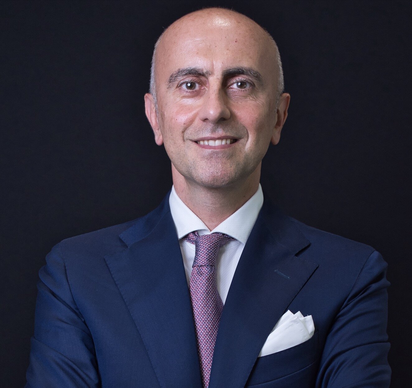
We use cookies to enable the functions required for this website, such as login or a shopping cart. You can find more information in our privacy policy.
Graduated in 1992 with a degree in dentistry from the University of Bologna, Italy, where he was subsequently adjunct professor from 1997 to 2000. Visiting researcher at the Department of Cariology, directed by Prof Ivo Krejci, at the University of Geneva between the years 2000 and 2004. Obtained a master’s degree in science (2003) and a PhD in dental materials (2005) at the University of Siena with Prof M. Ferrari. Researcher at the University of Bologna since 2006. Up to 2020, taught undergraduate courses on fixed prostheses on natural teeth and implants and prosthodontics at the University of Bologna. Italian Academy of Prosthetic Dentistry (AIOP) award winner for best research in the field of prosthodontics in the years 2005, 2008, and 2009. Obtained a PhD in material science in 2013 from the Faculty of Engineering, University of Bologna. Associate professor at the University of Modena and Reggio Emilia, teaching the courses of implant prosthodontics and removable prosthodontics in the school of dentistry. Dean of the master’s degree program in prosthodontics and implant prosthetics with advanced technologies, University of Bologna. Since 2004 collaborating with Prof G. Zucchelli in the development of clinical and research protocols related to implant-supported restorations, soft tissue management, and the interrelationship between prosthodontics and periodontics in the natural dentition. Active member of the AIOP and the Italian Academy of Osseointegration (IAO). Fellow of the International Team for Implantology since 2018.
1st Edition 2022 Book Hardcover; Two-volume book with slipcase; 21 x 28 cm, 1100 pages, 6557 illus Language: English Categories: Oral Surgery, Implantology Stock No.: 7736 ISBN 978-88-7492-091-4 QP Italy
International Journal of Computerized Dentistry, 1/2024
ScienceDOI: 10.3290/j.ijcd.b3963071, PubMed ID (PMID): 36928756Pages 27-35, Language: English, GermanMonaco, Carlo / Arena, Antonio / Marziali, Andrea / Consolo, Ugo
Aim: The present in vitro study aimed to evaluate the depth of reading of intraoral scanners (IOSs) within the gingival sulcus.
Materials and methods: A knife-edge preparation for a full crown was performed on a Frasaco model. The gingival sulcus of the scanned model was modified using a dedicated software program (Model Creator, exocad DentalCAD 2.4 Plovdiv) by setting the apical width (AW), coronal width (CW), and gingival sulcus depth (D). Two dental models with different gingival sulcus depths (1 or 2 mm) were printed using the digital light processing (DLP) technique. Each model was scanned 10 times. Seven different IOSs were used: Emerald, Trios 3, Carestream 3600, Dental Wings DWIO, CondorScan, True Definition Scanner (TDS), and Cerec Omnicam. Measurements of D values were performed using 3Shape 3D viewer software. The normality of the data distribution was evaluated using the Shapiro-Wilk test (P < 0.05). The nonparametric Levene’s test was used to check for homoscedasticity. The data were statistically analyzed using the Kruskal-Wallis test (α = 0.05) and the Nemenyi test.
Results: All IOSs were able to read within the 1-mm–deep gingival sulcus, albeit with some statistically significant differences (P < 0.001). TDS and Trios 3 were able to read within the 2-mm–deep gingival sulcus (P < 0.001).
Conclusions: The depth of reading of different IOSs can vary significantly. In the model with a 2-mm gingival sulcus, even in the absence of oral fluids, the depth of reading was incomplete, suggesting that deep preparations into the gingival sulcus are difficult to detect with IOSs.
Keywords: intraoral scanner (IOS), gingival sulcus, depth of reading, knife-edge preparation, digital light processing
The International Journal of Prosthodontics, 2/2023
DOI: 10.11607/ijp.7591, PubMed ID (PMID): 36445219Pages 228-232, Language: EnglishMonaco, Carlo / Scheda, Lorenzo / Arena, Antonio / di Fiore, Adolfo / Zucchelli, Giovanni
Purpose: To describe the treatment of malpositioned implants in the esthetic area using the angulated welded abutment (AWA) approach together with peri-implant soft tissue surgery.
Materials and Methods: A clinical case with extreme buccal angulation of two implants in the anterior maxilla was used to illustrate the AWA technique. After implant impression-taking, digital analysis was used to determine the ideal prosthetic angulation of the abutment and the ideal position of its screw hole in relation to the gingival margin of the adjacent teeth. The AWA was designed in two combinable components that were meant to be welded together. Before the welding process, an angulated screw was included in the abutment. Since the angulated screw was inside the abutment, the screwdriver hole could be designed as narrow as possible and put in an ideal position. After periodontal and peri-implant surgery were carried out, the AWA was applied to the implants.
Results: The AWA allowed correction of the prosthetic axis. Moreover, relocation of the screw hole allowed the gingival tissue to creep over the abutment. In this way, a new esthetic restoration can be placed after the mucogingival surgery.
Conclusion: The excessive misangulation of the implants was efficiently recovered. Further studies are needed to evaluate long-term clinical success, and standardization of this technique is required for routine clinical use.
Purpose: To describe the possible adverse effects of sodium hypochlorite (NaOCl) solutions, highconcentration alcohol solutions, and povidone-iodine products indicated for disinfection of inanimate surfaces against human coronavirus of the severe acute respiratory syndrome (SARS-CoV) on prosthesis materials, including zirconia, lithium disilicate, and acrylic resin.
Materials and Methods: A systematic literature research was conducted in the SCOPUS, PubMed/Medline, Web of Science, EMBASE, and Science Direct databases from January 2010 to February 2020 using a combination of the following MeSH/Emtree terms and keywords: “sodium hypochlorite”; “alcohol”; “ethanol”; “povidone-iodine”; “dental ceramic”; “zirconia”; “lithium disilicate”; and “acrylic resin”.
Results: A total of 538 studies were identified in the search during initial screening, 44 were subject to full-text evaluation, and 24 fulfilled the inclusion criteria. Seven articles on zirconia and lithium disilicate investigated the effect of NaOCl (0.5% and 1%), 96% isopropanol, and 80% ethanol on bond strength after saliva contamination. The remaining articles evaluated color alteration, surface roughness modifications, decrease in flexural strength, and bond strength of all cleaning agents on acrylic resin.
Conclusion: NaOCl solution (1%) for 1 minute is recommended to reduce SARSCoV infectivity and to minimize the risk of cross-contamination through prosthetic materials. An increase in surface roughness and color alteration on acrylic resin were recorded using 1% NaOCl, but without any clinical significance. A decrease in bonding strength was determined after using 1% NaOCl, 96% isopropanol, or 80% ethanol solutions on lithium disilicate. Silanization before the try-in procedure and the application of a second layer of silane after cleaning methods are recommended to improve the bond strength.
Purpose: To evaluate the influence of the crown-to-implant ratio (CI) on marginal bone loss (MBL) around short dental implants placed in the posterior mandible.
Materials and Methods: All patients treated with short implants (7-mm length) in the posterior mandible between 1994 and 2003 at the Dental Clinic of the Department of Neuroscience of the University of Padua (Italy) were retrospectively included in the analysis. MBL and clinical CI (cCI) were measured on the radiographs. Implant characteristics including implant diameter, prosthetic type, retention mode, antagonist type, veneering material, and implant surfaces were retrieved from local medical records. A generalized linear mixed model was estimated to identify the predictors of MBL.
Results: A total of 108 dental implants placed in 51 patients were included in the analysis. Mean follow-up was 16 years (range: 11 to 20 years). Mean cCI was 2.21 (SD = 0.31) with a mean crown height of 10.86 mm (SD = 0.99). Mean MBL was 1.42 mm (SD = 0.38). At multivariable analysis, cCI ≥ 2 was associated with higher MBL (regression coefficient: 0.27; 95% CI: 0.15 to 0.40), while implant characteristics, follow-up, and site were not associated with MBL. The effect of a cCI ≥ 2 was estimated in an increase of 0.28 mm in MBL (95% CI: 0.14 to 0.43 mm).
Conclusion: Higher cCI was associated with greater MBL of implant-supported fixed dental prostheses in short dental implants placed in the posterior mandible, while implant characteristics, follow-up, and site were not associated with MBL. However, the increase of 0.28 mm of MBL in patients with a cCI ≥ 2 may not be clinically relevant.
Keywords: crown-to-implant ratio, implant-supported dental prosthesis, marginal bone loss, prosthetic parameters, short implant
International Journal of Oral Implantology, 2/2018
PubMed ID (PMID): 29806668Pages 215-224, Language: EnglishZucchelli, Giovanni / Felice, Pietro / Mazzotti, Claudio / Marzadori, Matteo / Mounssif, Ilham / Monaco, Carlo / Stefanini, Martina
Purpose: To report the 5-year clinical and aesthetic outcomes of a novel surgical-prosthetic approach for the treatment of buccal soft tissue dehiscence around single dental implants.
Materials and methods: Twenty patients with buccal soft tissues dehiscence around single implants in the aesthetic area were treated by removing the implant-supported crown, reducing the implant abutment, coronally advanced flap in combination with connective tissue graft and final restoration. After the first year, patients were recalled three times a year until the final clinical re-evaluation performed 5 years after the final prosthetic crown. Complications, bleeding on probing (BoP), peri-implant probing depth (PPD), clinical attachment level (CAL), keratinized tissue height (KTH), soft tissue coverage and thickness (STT), patient satisfaction (VAS) and aesthetic assessment (PES/WES) were evaluated 5 years after the final restoration.
Results: Of the 20 patients enrolled in the study, 19 completed the study at 5 years. A total of 99.2% mean soft tissue dehiscence coverage, with 79% of complete dehiscence coverage, was achieved at 5 years. A statistically significant increase in buccal soft tissue thickness (0.3 mm 0.1-0.4 P < 0.001) and keratinized tissue height (0.5 mm 0.0-1.0; P < 0.001) at 5 years with respect to 1 year was demonstrated. The patient aesthetic evaluation showed high VAS scores with no statistical difference between 1 year and 5 years (8.75 ± 1.02 and 8.95 ± 0.91 respectively). A statistical significant PES/WES score improvement was observed between baseline and 5 years (9.48 ± 2.68; P < 0.001), but not between 1 and 5 years.
Conclusions: Successful aesthetic and soft tissue dehiscence coverage outcomes were well maintained at 5 years. The strict regimen of post-surgical control visits and the emphasis placed on the control of the toothbrushing technique could be critical for the successful long-term maintenance of soft tissue dehiscence coverage results.
Keywords: aesthetics, connective tissue, dental implant, mucogingival surgery, soft tissue dehiscence
Conflict-of-interest statement: The authors declare that they have no conflict of interest.
Purpose: Various materials and systems for bonding lithium disilicate to the tooth substrate are available to clinicians, who can adapt the materials to each clinical situation to maximize the performance of indirect esthetic restorations. This study aimed to evaluate the degree of conversion (DC) and the microhardness (MH) of a dual-curing and a light-curing cement under lithium disilicate discs of different thicknesses.
Materials and Methods: A total of 48 lithium disilicate (IPS e.max CAD, Ivoclar Vivadent) samples were prepared and divided into three groups (n = 16) according to the thickness (group A was 0.6 mm; group B was 1.0 mm; group C was 1.5 mm). Each group was further divided into two subgroups (n = 8) according to the resin cement employed, NX3 (Kerr) or Choice 2 (Bisco). A standardized quantity of cement was placed on the sample, and DC was evaluated with an attenuated total reflectance Fourier transformed infrared spectrophotometer (Nicolet IS10, Thermo Scientific). Twenty-four hours after DC was established, Vickers test was performed on the cement with a microindentometer (Leica Microsystems). Results were statistically analyzed with analysis of variance test and significance set at P < .05.
Results: Statistical analysis showed cement type had a significant influence (P = .005) on DC. MH results were influenced by thicknesses only between 0.6 and 1.5 mm when light-cured cement was employed.
Conclusion: The light-curing and the dualcuring cements reached comparable DCs between 0.6 and 1.5 mm. However, the light-curing resin showed a higher DC and MH.
Purpose: To evaluate the fracture strength and the failure mode of endodontically treated teeth restored with composite resin overlays with and without glass-fiber reinforcement.
Materials and Methods: A total of 32 extracted molars were divided into four equal groups. In the NFR-NFRC (no foundation restoration, no fiber-reinforced composite) and NFR-FRC (no foundation restoration, fiber-reinforced composite) groups, only a 5-mm-thick composite resin layer sealed the pulp chamber floors, whereas in the FR-NFRC (foundation restoration, no fiber-reinforced composite) and FR-FRC (foundation restoration, fiber-reinforced composite) groups, a 3.0-mm foundation restoration was used. NFR-NFRC and FR-NFRC groups were restored with composite resin overlays, whereas NFR-FRC and FR-FRC groups were restored with fiber-reinforced composite resin overlays. All specimens were subjected to mechanical loading in a computer-controlled masticator and then the fracture resistance was evaluated. Differences in means were compared using two-way ANOVA and Tukey's test. The level of significance was set at ɑ = 0.05.
Results: All specimens successfully completed the fatigue test. The least fracture-resistant group was NFR-FRC, exceeded by FR-NFRC, NFR-NFRC, and FR-FRC, in that order, with FR-FRC being the most fracture-resistant group. Statistically significant differences were detected between the pairs NFR-NFRC/FR-FRC (p = 0.001), NFR-FRC/FR-FRC (p = 0.001), and FR-NFRC/FR-FRC (p = 0.001). Eight vertical root fractures occurred in group FR-NFRC, six in group NFR-NFRC, four in group NFR-FRC, and none occurred in group FR-FRC.
Conclusions: Within the limitations of this in vitro study, the incorporation of glass fibers and the presence of a foundation restoration were found to increase the fracture resistance and can favorably influence the fracture mode.
Keywords: molars, resin-bonded onlays, root fractures
DOI: 10.3290/j.jad.a35251, PubMed ID (PMID): 26734682Pages 567-574, Language: EnglishMonaco, Carlo / Bortolotto, Tissiana / Arena, Antonio / Krejci, Ivo
Purpose: To evaluate the marginal adaptation and fracture load of composite resin onlays reinforced with different substructures.
Materials and Methods: Thirty-two extracted, caries-free premolars were selected for this study and endodontically treated. Group 1 was used as the control group, and the teeth were restored only with as-manufactured composite resin overlays. Group 2 teeth were restored with composite resin overlays with 3 fiber-reinforced composite (FRC) layers placed horizontally on the bottom of the restoration. Group 3 teeth were restored with composite resin overlays with 6 fiber-reinforced composite (FRC) layers placed as in group 2. Group 4 teeth were restored with composite resin overlays and FRC placed with an anatomical design. All specimens underwent SEM evaluation of their marginal adaptation before and after thermocycling and cyclic mechanical loading. All specimens were then subjected to a fracture test, recording the value for the initial (IF) and final (FF) failure. Differences in the means were compared using matched-pairs t-tests and one-way ANOVA. The level of significance was set at α = 0.05.
Results: No statistically significant difference between the four groups in terms of marginal adaptation was observed at the tooth/luting composite and luting composite/overlay interfaces before and after loading. The fracture loads of IF and FF, from most to least resistant were: group 4 (1431.8 ± 294.3 N / 1710.1 ± 326.6 N), group 3 (1428.1 ± 251.4 N / 1467.9 ± 242.4 N), group 2 (852.6 ± 413.5 N / 1058.1 ± 251.5 N) and group 1 (899.8 ± 352.7 N / 923.5 ± 318.8 N). Significant differences (p = 0.026) were observed comparing group 1 to groups 2 and 3, and group 1 to 4. Three irreparable fractures were found in group 3, four in group 2, and five in groups 1 and 4.
Conclusions: The presence or absence of reinforcement and the different configuration of the reinforcement fibers affect fracture strength but only partially the failure modality. The presence or absence of reinforcement does not alter marginal adaptation.
Keywords: FRC, endodontically treated teeth, glass fibers, fracture, marginal adaptation, onlays