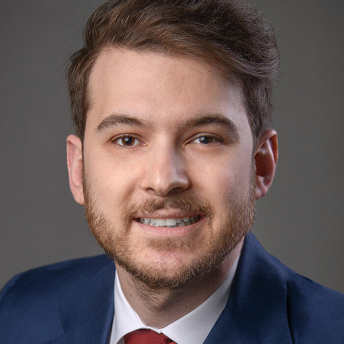
We use cookies to enable the functions required for this website, such as login or a shopping cart. You can find more information in our privacy policy.
Lorenzo Tavelli, DDS, MS, PhD isa full-time assistant professor and the director of the Postgraduate program in Periodontology at the Harvard School of Dental Medicine.
Dr. Tavelli graduated from the University of Milan and then completed his three-year residency in Periodontics and Master of Science at the University of Michigan. He is a diplomate of the American Board of Periodontology.
Dr. Tavelli’s main focus in clinical research and daily practice is microsurgical and minimally invasive soft tissue grafting procedures around teeth and dental implants, where he has been pioneering in the combined use of High Frequency Doppler Ultrasonography and wound healing biomarkers for assessing graft perfusion and the surgical outcomes. Dr. Tavelli has been extensively lecturing worldwide on these topics at the main scientific symposia. He has published more than 120 scholarly publications in the main international peer-reviewed journals while serving as an Associate Editor for Journal of Periodontal Research and Clinical Implant Dentistry and Related Research.
He was the recipients of several research awards, such as the 2024 ITI Andre Schroeder Research Prize, the 2023 American Academy of Periodontology (AAP) Earl Robinson Regeneration Award, the 2022 AAP Foundation Nevins Clinical Research Fellowship, the 2021 Goldman Clinical Research award from the Italian Society of Periodontology, and the 2020 AAP Balint Orban Research Award, among others.
DDS, MS, PhD isa full-time assistant professor and the director of the Postgraduate program in Periodontology at the Harvard School of Dental Medicine.
Dr. Tavelli graduated from the University of Milan and then completed his three-year residency in Periodontics and Master of Science at the University of Michigan. He is a diplomate of the American Board of Periodontology.
Dr. Tavelli’s main focus in clinical research and daily practice is microsurgical and minimally invasive soft tissue grafting procedures around teeth and dental implants, where he has been pioneering in the combined use of High Frequency Doppler Ultrasonography and wound healing biomarkers for assessing graft perfusion and the surgical outcomes. Dr. Tavelli has been extensively lecturing worldwide on these topics at the main scientific symposia. He has published more than 120 scholarly publications in the main international peer-reviewed journals while serving as an Associate Editor for Journal of Periodontal Research and Clinical Implant Dentistry and Related Research.
He was the recipients of several research awards, such as the 2024 ITI Andre Schroeder Research Prize, the 2023 American Academy of Periodontology (AAP) Earl Robinson Regeneration Award, the 2022 AAP Foundation Nevins Clinical Research Fellowship, the 2021 Goldman Clinical Research award from the Italian Society of Periodontology, and the 2020 AAP Balint Orban Research Award, among others.
Long term success in Regeneration3. Mar 2025 — 5. Mar 2025Guangzhou, China
Speakers: Ueli Grunder, Jaime Lozada, Lorenzo Tavelli, Istvan Urban
Osteology Foundation
The 14th International Symposium on Periodontics and Restorative Dentistry (ISPRD)
9. Jun 2022 — 12. Jun 2022Boston Marriott Copley Place, Boston, MA, United States of America
Speakers: Tara Aghaloo, Edward P. Allen, Evanthia Anadioti, Wael Att, Vinay Bhide, Markus B. Blatz, Scotty Bolding, Lorenzo Breschi, Jeff Brucia, Daniel Buser, Luigi Canullo, Daniele Cardaropoli, Stephen J. Chu, Donald Clem, Christian Coachman, Lyndon F. Cooper, Daniel Cullum, Lee Culp, José Carlos Martins da Rosa, Sergio de Paoli, Marco Degidi, Nicholas Dello Russo, Serge Dibart, Joseph P. Fiorellini, Mauro Fradeani, Stuart J. Froum, David Garber, Maria L. Geisinger, William Giannobile, Luca Gobbato, Ueli Grunder, Galip Gürel, Chad Gwaltney, Christoph Hämmerle, Robert A. Horowitz, Marc Hürzeler, David Kim, Gregg Kinzer, Christopher Köttgen, Ina Köttgen, Purnima S. Kumar, Burton Langer, Lydia Legg, Pascal Magne, Kenneth A. Malament, Jay Malmquist, George Mandelaris, Pamela K. McClain, Michael K. McGuire, Mauro Merli, Konrad H. Meyenberg, Craig M. Misch, Julie A. Mitchell, Marc L. Nevins, Myron Nevins, Michael G. Newman, Miguel A. Ortiz, Jacinthe M. Paquette, Stefano Parma-Benfenati, Michael A. Pikos, Giulio Rasperini, Pamela S. Ray, Christopher R. Richardson, Isabella Rocchietta, Marisa Roncati, Marco Ronda, Paul S. Rosen, Maria Emanuel Ryan, Irena Sailer, Maurice Salama, David M. Sarver, Takeshi Sasaki, Todd Scheyer, Massimo Simion, Michael Sonick, Sergio Spinato, Dennis P. Tarnow, Lorenzo Tavelli, Douglas A. Terry, Tiziano Testori, Carlo Tinti, Istvan Urban, Hom-Lay Wang, Robert Winter, Giovanni Zucchelli
Quintessence Publishing Co., Inc. USA
This author's journal articles
International Journal of Periodontics & Restorative Dentistry, Pre-Print
DOI: 10.11607/prd.7179, PubMed ID (PMID): 3905894426. Jul 2024,Pages 1-15, Language: EnglishUrban, Istvan A. / Di Martino, Maria / Rangel, Rodrigo / Latimer, Jessica / Forster, Andras / Tavelli, Lorenzo
A 45-year-old female patient presented with a lack of inter-implant papilla after a partially edentulous anterior area was rehabilitated with dental implants. The soft tissue phenotype and inter-implant papilla was augmented using the “iceberg” connective tissue graft, followed by a second surgical procedure where a strip gingival graft was combined with a connective tissue graft inserted underneath a pouch prepared into the previous “iceberg” connective tissue graft at the level of the crest (“garage” approach), further enhancing soft tissue volume in that region. This technique aims to improve mucosal thickness and supracrestal tissue height while addressing esthetic concerns associated with multiple implant placements in the anterior region. The final esthetic outcome was excellent, harmonious soft tissue with appropriate thickness, symmetry with adjacent teeth, well-shaped interdental and inter-implant papilla with high patient satisfaction, making this approach a valuable addition to a surgeon’s armamentarium. Future clinical studies are needed to evaluate the performance of this novel approach.
We report the successful treatment of multiple recession type (RT) 3 gingival recessions in periodontally compromised mandibular anterior teeth with limited keratinized tissue. A 35-yearold man with stage III, grade C periodontitis underwent a two-stage intervention. Initially, a modification of the connective tissue graft (m-CTG) wall technique was used as part of phenotype modification therapy. The CTG acted as a protective “wall,” securing space for periodontal regeneration, enhancing root coverage, soft tissue thickness, and keratinized mucosal width. Recombinant human fibroblast growth factor-2 and carbonate apatite promoted periodontal regeneration. This procedure successfully facilitated periodontal regeneration, resulting in the transition from RT3 to RT2 gingival recession and adequate keratinized mucosal width. Eighteen months later, the second surgery used a tunneled coronally advanced flap (TCAF) for root coverage. TCAF involved combining a coronally advanced flap and tunnel technique by elevating the trapezoidal surgical papilla and using a de-epithelialized CTG inserted beneath the tunneled flap. Root conditioning with ethylenediaminetetraacetic acid and enamel matrix derivative gel application were performed. Consequently, mean CAL gain was 5.3 mm, mean root coverage was 4.5 mm in height, and the gingival phenotype improved at the treated sites by the 12-month follow-up. This staged approach addresses the challenges of treating RT3 gingival recession with promising outcomes.
Keywords: RT3, gingival recession, case report, T-CAF, periodontal regeneration, connective tissue graft, CTG wall
Purpose: To conduct a systematic review of the existing literature, evaluating the different implant removal techniques in terms of success rate, complications and implant site management. Materials and methods: Two independent reviewers conducted an electronic literature search in PubMed/Medline, Web of Science, Scopus and EBSCO databases as well as a manual search to identify eligible clinical studies up to August 12, 2024. The protocol was designed in accordance with the Preferred Reporting Items for Systematic Reviews and Meta- Analyses (PRISMA) Statement. Results: The initial electronic search resulted in 1076 studies. The systematic application of inclusion and exclusion criteria resulted in 45 articles that met the purpose of the study. A sample of 736 patients and 1381 implants removed was obtained from the selected articles. Peri-implantitis was the most common reason for explantation (79.3%), and twelve implant removal devices or techniques were identified. Complications associated with implant removal were reported in a total of 5 studies, including 16 implants (1.2%). Conclusions: Within the limitations of this systematic review, it can be established that in subjects requiring the removal of osseointegrated endosseous dental implants, the current removal techniques are very effective in terms of success and low complication rates. However, site management after implant removal was poorly reported.
Keywords: dental implants, osseointegration, peri-implantitis, device removal, alveolar bone loss, titanium
International Journal of Periodontics & Restorative Dentistry, Pre-Print
DOI: 10.11607/prd.7476, PubMed ID (PMID): 397757147. Jan 2025,Pages 1-20, Language: EnglishUrban, Istvan A. / Mancini, Leonardo / Akhondi, Samuel / Tavelli, Lorenzo
It is well known that keratinized mucosa (KM) plays a crucial role for maintaining peri implant health and esthetic outcomes. The Strip Gingival Graft (SGG) technique, which involved an apically positioned flap (APF), in combination with an autogenous SGG and a xenogeneic collagen matrix (XCM), demonstrated its efficacy in re-establishing an adequate amount of KM width at implant sites. Nevertheless, it is still unclear whether harvesting the SGG from the palate (pSGG) or from the buccal aspect of natural dentition (bSGG) affects the esthetic outcomes at the augmented implant sites. Therefore, the objective of the present study was to compare the esthetic outcomes of dental implants augmented with either bSGG + XCM or pSGG + XCM. The present study was designed as a single-center retrospective study, assessing the esthetic and colorimetric outcomes of peri-implant KM augmentation with either pSGG + XCM or bSGG + XCM in cohort of 49 subjects. The two groups were compared in terms of colorimetric outcomes, assessed on clinical photograph using specific software able to identify and quantify the predominant color within the peri implant soft tissue. Colorimetric comparisons with adjacent untreated sites were also investigated. In addition, the Pink Esthetic Score (PES) and the Subjective Esthetic Score (SEE) were performed to further assess the esthetic outcomes of pSGG + XCM and bSGG + XCM. The colorimetric analysis did not show statistically significant differences among sites augmented with pSGG + XCM, sites augmented with bSGG + XCM, and untreated sites. Implants treated with bSGG + XCM showed significantly greater PES (in terms of shape of the mesial and distal papilla, level of the soft tissue margin, soft tissue contour, anatomy of the alveolar process, and final PES) and SEE compared to implants augmented with pSGG + XCM. The present study demonstrated that implant sites augmented with APF with either pSGG + XCM or bSGG + XCM did not show different colorimetric outcomes compared to adjacent untreated sites, while bSGG + XCM obtained superior professional and subjective esthetic scores compared to pSGG + XCM.
International Journal of Periodontics & Restorative Dentistry, Pre-Print
DOI: 10.11607/prd.7197, PubMed ID (PMID): 3882027831. May 2024,Pages 1-22, Language: EnglishZwanzig, Kai / Akhondi, Samuel / Tavelli, Lorenzo / Lanis, Alejandro
Introduction: The presence of adequate keratinized mucosa (KM) around dental implants and natural
dentition is pivotal for the long-term success of dental restorations. Despite various techniques to
augment KM, challenges persist in achieving stable, keratinized, and adherent mucosa, especially in
the context of significant muscle pull or compromised tissue conditions. This study introduces a novel
application of titanium pins for the fixation of free gingival grafts (FGG) and apically repositioned
flaps (APF) during vestibuloplasty, aiming to overcome important limitations associated with
traditional suturing methods and shorten the treatment time and patient morbidity. Methods: Three
patients with insufficient KM width, presenting discomfort during oral hygiene and inflammation
around implant restorations and natural teeth, underwent soft tissue augmentation using titanium pins
traditionally used in guided bone regeneration (GBR) for the stabilization of FGGs and APFs. This
method ensures intimate contact between the graft and the periosteum, facilitating proper graft
perfusion and revascularization, minimizing shrinkage and the risk for necrosis of the graft. Results:
Postoperative follow-up revealed successful integration of the grafts, with minimal shrinkage and
increased width and depth of KM. The use of titanium pins allowed for reliable fixation in challenging
surgical sites, where traditional suturing methods were impractical due to the presence of extensive
muscle pull and an unstable recipient bed. Conclusion: The application of titanium pins for the fixation
of FGGs and APFs during vestibuloplasty provides a promising alternative to traditional suturing
techniques, particularly in complex cases where the recipient bed is suboptimal for suturing. This
method simplifies and shortens the procedure, offering a predictable outcome with increased
mechanical stability and minimal shrinkage of the graft. Randomized clinical trials are recommended
to further evaluate the efficacy of this technique.
Keywords: Free Gingival Graft, Dental Implants , Apically repositioned flap, Titanium pins, Vestibuloplasty, Graft Survival, Periodontal Surgery, Titanium Pins, Transplants
The peri-implant mucosal barrier “seal” plays a significant role in maintaining peri-implant health, but its efficacy in the presence of inflammation is lower than that of natural teeth due, primarily, to the absence of collagen fiber insertion into the implant/abutment surface. To test the influence of cementum upon collagen fiber insertion/orientation after tooth removal, a customized root-cementum abutment was fabricated using a natural tooth root fragment. For that, an extracted root fragment, preserving both cementum and periodontal ligament, was cemented to the titanium abutment and used as a healing abutment of an immediate implant placed into the fresh extraction socket. Three months after implant placement, firm resistance to probing was noted clinically upon follow-up evaluation and histological and FE-SEM analyses confirmed perpendicular collagen fiber embedding into the root-cementum abutment surface. This proof-of-concept unveils the role of cementum on fiber insertion/orientation and sheds light on the relevance of enhancing the sealing of the peri-implant mucosal barrier to protect the underlying bone by utilizing a customized abutment that allows for the insertion of connective tissue fibers.
Keywords: dental implants, collagen fiber, connective tissue, integration, case reports
Soft tissue augmentation procedures are often needed at anterior dental implants to address esthetic concerns. The surgical techniques for vertical soft tissue augmentation and papilla augmentation at implant sites, their predictability, and outcomes have recently gained popularity among clinicians to meet patients’ high esthetic demands. This study describes the application of the tunneled coronally advanced flap for vertical soft tissue reconstruction and papilla augmentation (verTCAF) for the treatment of peri-implant soft tissue dehiscences (PSTDs) characterized by papilla loss. Twelve patients with isolated PSTD in the anterior region were consecutively enrolled and treated with the verTCAF in combination with a connective tissue graft. This approach required a temporary phase prior to the surgery. The verTCAF involved a horizontal and vertical incision, with the opening of one surgical papilla one tooth more distal than the implant site. The anterior region, including the implant site and the interproximal soft tissue were tunneled. A connective tissue graft (CTG) was inserted and stabilized from the lateral opening, where the surgical papilla was raised. After the soft tissue augmentation and a new temporary phase of at least 3-4 months, the cases were finalized with new restorations. Patient- reported outcomes, and clinical and ultrasonographic measures were collected over 1 year to assess the results of the procedure. One year after the intervention, the subjects reported a significant improvement in the esthetics (93.92 visual analogue scale points at the last visit). The overall percentage of black triangle reduction, compared to baseline, was 83.3%. The mean PSTD coverage and complete PSTD coverage were 92.78% and 66.7%, respectively. The average mesial and distal papilla gains were 1.79 mm and 1.46 mm, respectively. A significant increase of the peri-implant soft tissue phenotype parameters was observed, together with a substantial root coverage and papilla gain in natural dentition. The present case series described the application and the promising outcomes of the verTCAF + CTG for PSTDs, exhibiting papilla loss and adjacent teeth with midfacial and interproximal recession.
This split-mouth trial investigated the efficacy of treating bilateral gingival recessions with either a xenogeneic cross-linked collagen matrix (CCM), or recombinant human platelet derived growth factor (rhPDGF-BB) with a bone allograft (AG). Ten patients were treated with the coronally advanced flap (CAF), either with a CCM, or rhPDGF-BB + AG. The primary outcome was percentage of mean root coverage (mRC) at 12 months. Additional outcomes included clinical, volumetric, patient-reported outcome measures (PROMs) and ultrasonographic assessment of gingival thickness (GT) and position of the buccal bone (uBD). At 12 months, both groups showed significant improvements, with a mRC of 78.6% in the CCM group, and 82.3% for the rhPDGF-BB + AG sites. 3D analysis of both groups showed comparable volumetric gain. CCM-treated sites displayed higher ultrasonographic echogenicity in GT (p<.01) than rhPDGF-BB + AG sites. The rhPDGF-BB + AG group showed greater reduction in the buccal bone dehiscence (mean 2.03 mm, p<0.01), less swelling during the first three days, and slighty greater mean root coverage. CCM and rhPDGF-BB + AG showed to be effective in the treating multiple adjacent gingival recessions. CCM promotes greater gain in gingival thickness, while rhPDGF-BB + AG resulted in a significantly less buccal bone dehiscense.
Keywords: Gingival Recession, Root Coverage, Regeneration, Ultrasonography, Periodontics, Growth factors, Evidence-Based Dentistry, Platelet-derived growth factor, PDGF
The integrity and phenotype of periodontal soft tissues significantly influence the outcome of surgical periodontal regenerative therapy. In cases with thin gingival phenotype, treating infrabony defects surgically can worsen gingival recession and loss of papillae. This report outlines a surgical approach for addressing infrabony defects at sites with gingival recession and thin phenotype. The treatment involves using a tunneled coronally advanced flap (TCAF) to obtain access for defect debridement, root instrumentation, graft placement, and tissue advancement for root coverage. A connective tissue graft (CTG) is secured to the two teeth flanking the infrabony defect with two subperiosteal sling (SPS) sutures to create a buccal soft tissue wall and to tent up the papilla overlying the defect to provide and maintain the necessary space for biomaterials and clot stability. The treatment significantly improved interproximal clinical attachment levels, tissue phenotype, and root coverage one-year post surgery. Treatment outcomes suggest that this approach may be used to effectively treat isolated infrabony defects associated with gingival recession.
Keywords: connective tissue graft, osseous defects, periodontal diseases, periodontal regeneration
Purpose: To conduct a comprehensive umbrella review to synthesise existing evidence and critically evaluate the significance of keratinised mucosa width in peri-implant health and assess the consistency and heterogeneity among previous systematic reviews on this topic. Materials and methods: A comprehensive search strategy was implemented across multiple databases. Eligible studies were screened and data were extracted. Methodological quality was assessed using A MeaSurement Tool to Assess systematic Reviews version 2, and strength of evidence was evaluated using the Grading of Recommendations Assessment, Development and Evaluation criteria. A meta-meta-analysis using Hedges’ g as the effect size measure was performed to investigate the outcomes of implant therapy in patients with (control) and without adequate keratinised mucosa width (case). Results: Ten systematic reviews, published between 2012 and 2023, were included. Significant effect sizes were found for mucosal recession, Gingival Index/modified Gingival Index, modified Plaque Index and marginal bone loss. Specifically, narrow keratinised mucosa width ( 2 mm) was associated with increased mucosal recession (equivalent odds ratio 4.05, P = 0.03), higher Gingival Index/modified Gingival Index scores (equivalent odds ratio 3.131, P = 0.001), elevated modified Plaque Index scores (equivalent odds ratio 5.34, P = 0.005) and greater marginal bone loss (equivalent odds ratio 1.852, P = 0.0007). No significant associations were observed for bleeding on probing, pocket depth changes or pocket depth values. Follow-up time did not have a significant effect on these outcomes. Conclusions: Inadequate keratinised mucosa width ( 2 mm) correlated with increased mucosal recession, higher Gingival Index/modified Gingival Index, Plaque Index/modified Plaque Index scores and greater marginal bone loss. However, there is still a lack of sufficient evidence indicating the impact on bleeding on probing, pocket depth, implant survival and disease prevalence (no significant association or insufficient evidence).
Keywords: keratinised mucosa, keratinised tissue, peri-implant health, peri-implant mucositis, peri-implantitis
The authors declare there are no conflicts of interest relating to this study.