Utilizamos cookies para habilitar las funciones necesarias para este sitio web, como el inicio de sesión o la cesta de la compra. Puede encontrar más información en nuestra política de privacidad.
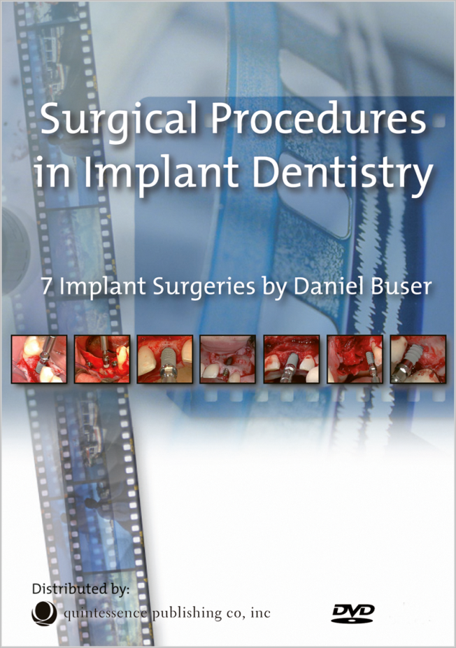
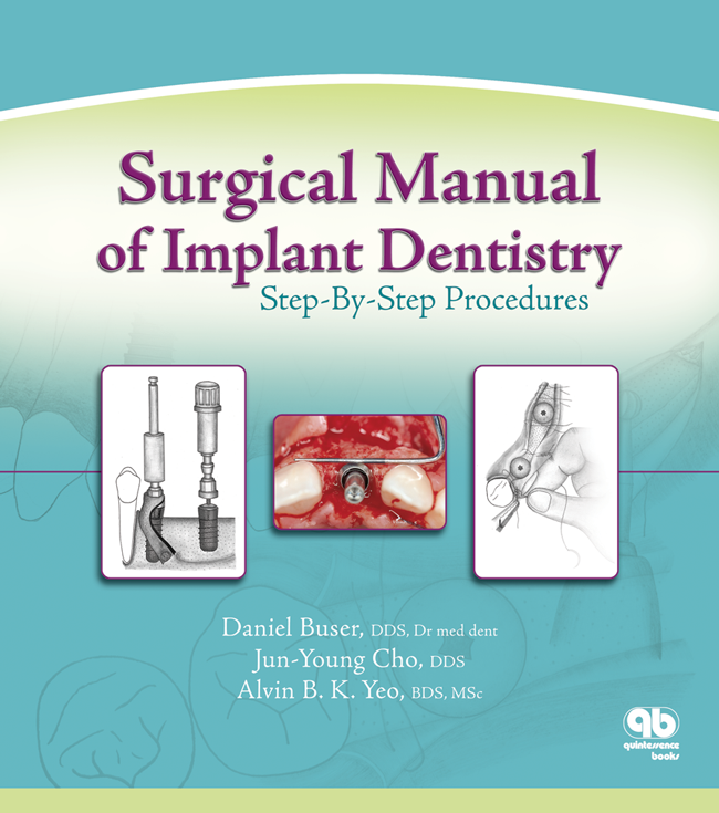
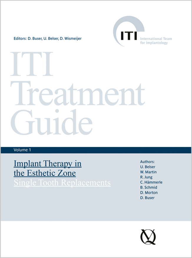
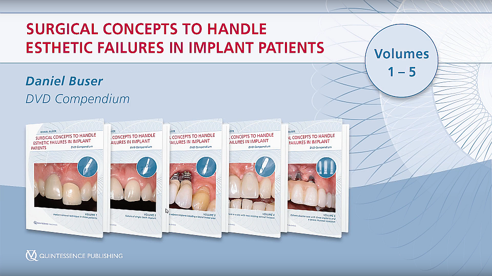
Daniel Buser, DDS, Prof em Dr med dent, is one of the most experienced international implant surgeons, with 35 years of surgical experience. He is past professor and chairman in the Department of Oral Surgery at the University of Bern in Switzerland and past president of the European Association for Osseointegration, the Swiss Society of Oral Implantology, the Swiss Society of Oral Surgery and Stomatology, and the International Team for Implantology (ITI). He is the recipient of numerous awards, including the ITI Honorary Fellowship and the Morton Amsterdam Award. He has authored and coauthored over 400 publications and is editor of 30 Years of Guided Bone Regeneration (Quintessence, 2021), which was recently published in its third edition. Dr Buser is cofounder of the Buser & Sculean Academy, which offers continuing education courses in periodontology and implant dentistry, and lectures nationally and internationally.
2nd revised and expanded Edition 2025 Libro Hardcover, 21 x 28 cm, 352 páginas, 1040 ilustración Idioma: Alemán Categorías: Implantología, Cirugía oral ISBN 978-3-86867-587-0 QP Deutschland
3rd Edition 2022 Libro Hardcover, 21,6 x 28 cm, 344 páginas, 1040 ilustración Idioma: Inglés Categoría: Implantología Nº de stock: B8038 ISBN 978-0-86715-803-8 QP USA
Aktuelle Therapievarianten und Materialien für Einzelzahnersatz
Serie: ITI Treatment Guide Series, Band 10 1. Auflage 2018 Libro Hardcover, 21 x 28 cm, 444 páginas, 1340 ilustración Idioma: Alemán Categoría: Implantología ISBN 978-3-86867-382-1 QP Deutschland
1st Edition 2017 DVD 5 DVDs in a box; NTSC/PAL; runtime 120 min Idioma: Inglés Categorías: Odontología estética, Implantología, Cirugía oral Nº de stock: C0962 ISBN 978-1-78698-011-3 QP Deutschland
Serie: ITI Treatment Guide Series, Band 09 1. Auflage 2017 Libro Hardcover, 21 x 28 cm, 312 páginas, 536 ilustración Idioma: Alemán Categoría: Implantología ISBN 978-3-86867-317-3 QP Deutschland
Serie: ITI Treatment Guide Series, Band 07 1. Auflage 2014 Libro Hardcover, 232 páginas, 600 en color Idioma: Alemán Categoría: Implantología ISBN 978-3-86867-235-0 QP Deutschland
Details make perfectionoctubre 24, 2024 — octubre 26, 2024MiCo - Milano Convention Centre, Milano, Italia
Ponentes: Bilal Al-Nawas, Gil Alcoforado, Federico Hernández Alfaro, Sofia Aroca, Wael Att, Gustavo Avila-Ortiz, Kathrin Becker, Anne Benhamou, Juan Blanco Carrión, Dieter Bosshardt, Daniel Buser, Francesco Cairo, Paolo Casentini, Raffaele Cavalcanti, Tali Chackartchi, Renato Cocconi, Luca Cordaro, Luca De Stavola, Nuno Sousa Dias, Egon Euwe, Vincent Fehmer, Alberto Fonzar, Helena Francisco, Lukas Fürhauser, German O. Gallucci, Oscar Gonzalez-Martin, Dominik Groß, Robert Haas, Alexis Ioannidis, Simon Storgård Jensen, Ronald Jung, France Lambert, Luca Landi, Georg Mailath-Pokorny jun., Silvia Masiero, Iva Milinkovic, Carlo Monaco, José Nart, José M. Navarro, Katja Nelson, Manuel Nienkemper, David Nisand, Michael Payer, Sergio Piano, Bjarni E. Pjetursson, Sven Reich, Isabella Rocchietta, Giuseppe Romeo, Irena Sailer, Mariano Sanz, Ignacio Sanz Martín, Frank Schwarz, Shakeel Shahdad, Massimo Simion, Ralf Smeets, Benedikt Spies, Bogna Stawarczyk, Martina Stefanini, Hendrik Terheyden, Tiziano Testori, Daniel Thoma, Ana Torres Moneu, Piero Venezia, Lukas Waltenberger, Hom-Lay Wang, Stefan Wolfart, Giovanni Zucchelli, Otto Zuhr
European Association for Osseintegration (EAO)
The Buser & Belser Master Course on Esthetic Implant Dentistry
Ponentes: Samir Abou-Ayash, Urs C. Belser, Dieter Bosshardt, Daniel Buser, Jordi Caballé-Serrano, Stephen Chen, Vincent Fehmer, Manrique Fonseca, Ronald Jung, Irena Sailer, Anton Sculean
Buser & Sculean Academy
The 14th International Symposium on Periodontics and Restorative Dentistry (ISPRD)
junio 9, 2022 — junio 12, 2022Boston Marriott Copley Place, Boston, MA, Estados Unidos de América
Ponentes: Tara Aghaloo, Edward P. Allen, Evanthia Anadioti, Wael Att, Vinay Bhide, Markus B. Blatz, Scotty Bolding, Lorenzo Breschi, Jeff Brucia, Daniel Buser, Luigi Canullo, Daniele Cardaropoli, Stephen J. Chu, Donald Clem, Christian Coachman, Lyndon F. Cooper, Daniel Cullum, Lee Culp, José Carlos Martins da Rosa, Sergio De Paoli, Marco Degidi, Nicholas Dello Russo, Serge Dibart, Joseph P. Fiorellini, Mauro Fradeani, Stuart J. Froum, David Garber, Maria L. Geisinger, William Giannobile, Luca Gobbato, Ueli Grunder, Galip Gürel, Chad Gwaltney, Christoph Hämmerle, Robert A. Horowitz, Marc Hürzeler, David Kim, Gregg Kinzer, Christopher Köttgen, Ina Köttgen, Purnima S. Kumar, Burton Langer, Lydia Legg, Pascal Magne, Kenneth A. Malament, Jay Malmquist, George Mandelaris, Pamela K. McClain, Michael K. McGuire, Mauro Merli, Konrad H. Meyenberg, Craig M. Misch, Julie A. Mitchell, Marc L. Nevins, Myron Nevins, Michael G. Newman, Miguel A. Ortiz, Jacinthe M. Paquette, Stefano Parma-Benfenati, Michael A. Pikos, Giulio Rasperini, Pamela S. Ray, Christopher R. Richardson, Isabella Rocchietta, Marisa Roncati, Marco Ronda, Paul S. Rosen, Maria Emanuel Ryan, Irena Sailer, Maurice Salama, David M. Sarver, Takeshi Sasaki, Todd Scheyer, Massimo Simion, Michael Sonick, Sergio Spinato, Dennis P. Tarnow, Lorenzo Tavelli, Douglas A. Terry, Tiziano Testori, Carlo Tinti, Istvan Urban, Hom-Lay Wang, Robert Winter, Giovanni Zucchelli
Quintessence Publishing Co., Inc. USA
Artículos de este autor en revistas
The International Journal of Prosthodontics, Pre-Print
DOI: 10.11607/ijp.7684, ID de PubMed (PMID): 36165882septiembre 22, 2022,Páginas 1-8, Idioma: InglésEl Kholy, Karim / Doliveux, Simon / Mattheos, Nikos / Buser, Daniel
This case report describes a novel approach combining orthodontic and implant digital treatment planning workflows to enable prosthetically driven implant placement before orthodontic treatment is performed or completed. The computer-assisted implant placement is performed using a static surgical guide based on the future positions of the teeth after completion of orthodontic treatment. This innovative approach allows for select partially edentulous patients to undergo fixed prosthetic rehabilitation before orthodontic treatment is completed.
International Journal of Periodontics & Restorative Dentistry, 4/2024
DOI: 10.11607/prd.6626, ID de PubMed (PMID): 37819849Páginas 434-443, Idioma: InglésFonseca, Manrique / Molinero-Mourelle, Pedro / Dönmez, Mustafa Borga / Abou-Ayash, Samir / Buser, Daniel / Sculean, Anton / Yilmaz, Burak
Dental implants are commonly used to replace missing single teeth. However, esthetic rehabilitation of an adjacent tooth may also be required due to diastemas, crowding, or existing large direct restorations to improve the final esthetic outcome. With the advancements in ceramics and bonding techniques, minimally invasive esthetic approaches have become viable for compromised spacing issues. This case report describes a dental technique for the esthetic rehabilitation of compromised anterior spacing with a customized zirconia implant abutment at a maxillary central incisor site and a partial ceramic veneer bonded to the adjacent central incisor.
Palabras clave: Anterior spacing; case report; implants; partial laminate veneer; prosthetic dentistry
This case report describes a novel approach combining orthodontic and implant digital treatment planning workflows to enable prosthetically driven implant placement before orthodontic treatment is performed or completed. The computer-assisted implant placement is performed using a static surgical guide based on the future positions of the teeth after completion of orthodontic treatment. This innovative approach allows for select partially edentulous patients to undergo fixed prosthetic rehabilitation before orthodontic treatment is completed.
Purpose: To analyze the effect of implant placement and loading protocols (protocol types) on the survival of single implant tooth replacements in different locations.
Materials and methods: An electronic search was conducted to identify clinical trials regarding outcomes of single implants subjected to different treatment protocols. A weighted mean survival rate for each protocol type in the anterior maxilla, anterior mandible, posterior maxilla, and posterior mandible was calculated. Study design, sample size, and outcome homogeneity were used to evaluate the validation of each protocol type in different locations.
Results: A total of 45 publications (13 RCTs, 21 prospective studies, and 11 retrospective studies) were included. The anterior maxilla was the most reported site (35 studies, 1,391 implants, weighted survival rate: 97.5% to 99.6%). Immediate placement + conventional loading (Type 1C) and late placement + immediate restoration/loading (Type 4A) were scientifically and clinically validated (SCV). For the posterior maxilla (19 studies, 567 implants, weighted survival rate: 85.7% to 100%), Type 1C was SCV. The anterior mandible was the least-reported site (three studies, 42 implants, weighted survival rate: 98.5% to 100%). For the posterior mandible (13 studies, 447 implants, weighted survival rate: 95.0% to 100%), late placement + conventional loading (Type 4C) was SCV. It was not possible to perform a metaanalysis due to the limited number of controlled studies that had the same comparison and considerable heterogeneity in study design.
Conclusion: Differences were found in the level of scientific evidence between the anterior and posterior and the maxilla and mandible, indicating that location is a consideration when selecting treatment protocol for a single implant.
Palabras clave: dental implants, placement and loading protocol, single implant
Die Implantattherapie bei älteren Menschen bedarf einer gezielten Planung. Dabei spielt die Unterscheidung zwischen dem dritten und vierten Lebensabschnitt für die Behandlungsplanung und die anschließende zahnärztliche Betreuung eine wichtige Rolle, da diese Patientengruppen sich fundamental voneinander unterscheiden. Für Menschen des dritten Lebensabschnitts steht das gesamte Spektrum der zahnärztlichen Implantologie zur Verfügung, allerdings müssen chronische Erkrankungen, Medikation und manuelle Geschicklichkeit beachtet werden. Da dentale Implantate meist deutlich länger als 20 Jahre im Mund verbleiben, muss prospektiv die zukünftige Abnahme der biologischen, sozialen und kognitiven Funktionen bei der Planung berücksichtigt werden. Im vierten Lebensabschnitt ist das Leben von der Abhängigkeit bei den Aktivitäten des täglichen Lebens (ADL), Multimorbidität, Polypharmazie und Institutionalisierung bestimmt. Es gibt fast keine wissenschaftliche Evidenz zur Implantattherapie dieser Patientengruppe. Die wenigen vorhandenen Studien zeigen eine sehr hohe Implantatüberlebensrate und es gibt Hinweise auf verringerte akute periimplantäre Entzündungsreaktionen. Gerade Patienten in der vierten Lebensphase könnten am meisten vom funktionellen Gewinn einer implantatstabiliserten Totalprothese profitieren, wenn die muskulären Fähigkeiten zur Stabilisierung einer Prothese nachlassen. Allerdings müssen Handhabung, Nachsorge und tägliche Pflege gesichert werden, bevor mit einer solchen Therapie im Sinne des Nichtschadensgebots begonnen wird. Menschen im palliativen Kontext weisen zunehmend Implantate auf, die die Betroffenen und die Pflegenden vor große Herausforderungen stellen können. Hier müssen die Implantatprothesen rechtzeitig „rückgebaut“ werden, um Schmerzen und Infektionen zu vermeiden.
Manuskripteingang: 06.01.2021, Annahme: 19.02.2021
Palabras clave: Implantologie, Gerodontologie, dritter Lebensabschnitt, vierter Lebensabschnitt, chirurgische Strategien, prothetische Strategien
The International Journal of Oral & Maxillofacial Implants, 5/2020
DOI: 10.11607/jomi.8045, ID de PubMed (PMID): 32991653Páginas 1013-1020, Idioma: InglésHicklin, Stefan Paul / Janner, Simone Fm / Schnider, Nicole / Chappuis, Vivianne / Buser, Daniel / Brägger, Urs
Purpose: The hydrophilic implant surface (INICELL) is a chemical alteration of a sandblasted and thermally acid-etched surface that should lead to long-term osseointegration. This study investigated 3-year results after early loading of implants with a hydrophilic, moderately rough surface in occlusal contact.
Materials and Methods: This prospective case series study was conducted in subjects with partially edentulous mandibles. Implants were placed on day 21 and loaded with a provisional reconstruction after at least 21 days of healing (baseline, day 0) if their implant stability quotient (ISQ) was ≥ 70 (mean of three measurements) and were replaced by definitive porcelain-fused-to-metal prostheses at the 6-month follow-up visit. Follow-up examinations were planned 1, 3, 6, 12, and 36 months after baseline.
Results: A total of 20 implants were placed in 15 patients (mean age: 51 years, range: 32 to 67 years). After 36 months, all implants were osseointegrated, and no suppuration was recorded. Small changes of bone level were observed between 3 months and 36 months. At 36 months, the median values of the 20 implants were 0.25 (range: 0 to 0.5, SD: 0.17), 0.25 (range: 0 to 1, SD: 0.27), and 4 (range: 2 to 7.25, SD: 1.17) for the mean modified Plaque Index (mPI), mean modified Sulcus Bleeding Index (mSBI), and mean probing pocket depth, respectively. The pairwise analysis between 3 and 36 months showed an improvement in the mean mPI (P = .0126) and mean mSBI (P = .0059). After 36 months, all patients (n = 15) were fully satisfied with a mean of 9.43 (range: 8 to 10, SD: 0.678) at the visual analog scale.
Conclusion: Early functional loading of implants with a hydrophilic, moderately rough outer surface in occlusal contact 21 days after healing appears to be a safe and feasible treatment option when placed in the posterior mandible of partially edentulous patients.
Palabras clave: delayed placement, dental implants, early loading, hydrophilic implant surface
Peri-implantitis is a biofilm-mediated inflammatory process that leads to soft- and hard-tissue breakdown. Periimplant diseases are the most frequent biologic implant complication in daily practice. Certain systemic conditions as well as detrimental habits have been demonstrated to negatively impact peri-implant tissue health and stability. In addition, several local predisposing factors were also identified to be associated with the development of periimplantitis, such as the lack of keratinized mucosa, residual cement or a micro-rough implant surface exposed to the oral cavity or the peri-implant sulcus. This clinical review paper will focus on the latter risk factor, which is often caused by poor surgical performance by the clinician during implant surgery.
Palabras clave: Peri-implantitis, peri-implant disease, dental implant, implant infection, risk factor, surgical risk factors
The International Journal of Oral & Maxillofacial Implants, 6/2019
DOI: 10.11607/jomi.7657, ID de PubMed (PMID): 31532826Páginas 1328-1336a, Idioma: InglésMonje, Alberto / Chappuis, Vivianne / Monje, Florencio / Muñoz, Fernando / Wang, Hom-Lay / Urban, Istvan A. / Buser, Daniel
Purpose: There is a lack of knowledge concerning the critical buccal bone thickness required for securing favorable functional and esthetic outcomes, conditioned to the dimensional changes after implant placement. A preclinical study was therefore carried out to identify the critical buccal bone wall thickness for minimizing bone resorption during physiologic and pathologic bone remodeling.
Materials and Methods: A randomized, two-arm in vivo study in healthy beagle dogs was carried out. The first group of dogs was sacrificed 8 weeks after implant placement for histomorphometric examination of postsurgical resorption of the buccal bone wall. The second group of dogs was monitored during three ligature-induced peri-implantitis episodes and a spontaneous progression episode. Morphometric and clinical variables were defined for the study of physiologic and pathologic buccal and lingual bone loss.
Results: Seventy-two implants were placed in healed mandibular ridges of 12 beagle dogs. Two groups were defined: 36 implants were placed in sites with a thin buccal bone wall ( 1.5 mm), and 36 were placed in sites with a thick buccal bone wall (≥ 1.5 mm). No implants failed during the study period. For the great majority of the histomorphometric parameters, a critical buccal bone wall thickness of at least 1.5 mm seemed to be essential for maintaining the buccal bone wall during physiologic and pathologic bone resorption. Suppuration (+) and mucosal recession (-) were more often associated with implants placed in sites with a thin buccal bone wall.
Conclusion: A critical buccal bone wall thickness of 1.5 mm at implant placement is advised, since a thicker peri-implant buccal bone wall (≥ 1.5 mm) is exposed to significantly less physiologic and pathologic bone loss compared with a thinner buccal bone wall ( 1.5 mm).
Palabras clave: alveolar bone, dental implants, diagnostic, implant stability, peri-implant mucositis, peri-implantitis
Over the past decade, the use of digital technology in implant treatment planning and static computer-assisted implant surgery (sCAIS) has revolutionized the planning and execution of guided implant surgeries. However, the predictability of achieving a high degree of accuracy when using a digital workflow and sCAIS has been a subject of debate. For sCAIS procedures to transition from clinical success in individual cases to a broadly applicable procedure, a better understanding and control of variables that affect their accuracy is essential. Recently, a research team in the Department of Oral Surgery and Stomatology at the University of Bern launched a series of in-vitro investigations to further analyze the impact and magnitude of potential variables involved in the digital treatment planning of sCAIS procedures that can have a significant effect on the accuracy of sCAIS. This article presents the rationale and summary of their findings.
Palabras clave: Computer-assisted, implant surgery, guided surgery, surgical guides