Regenerative Measures for Osseous Defect Repair and Optimal Esthetics
Regenerative Measures for Osseous Defect Repair and Optimal Esthetics
Category: Periodontics
Language(s): English, German
Publication year: 2006
Video source: APW DVD Journal
Series: APW DVD Journal
Procedure
Theoretical Part
- Adult male with a deep and broad intraosseous bone defect located on tooth #13
- The indication for modified papilla preservation in the scope of regenerative therapy was established based on the width of the diastema
- Regenerative periodontal therapy with Emdogain and a Bio-Oss® cancellous bone graft
- Emdogain is applied to the root surface to stimulate regeneration of periodontal structures
- To prevent graft collapse and to minimize the risk of development of too large a recession in this esthetically important region, the defect was filled with Bio-Oss® cancellous bone material
Practical Part
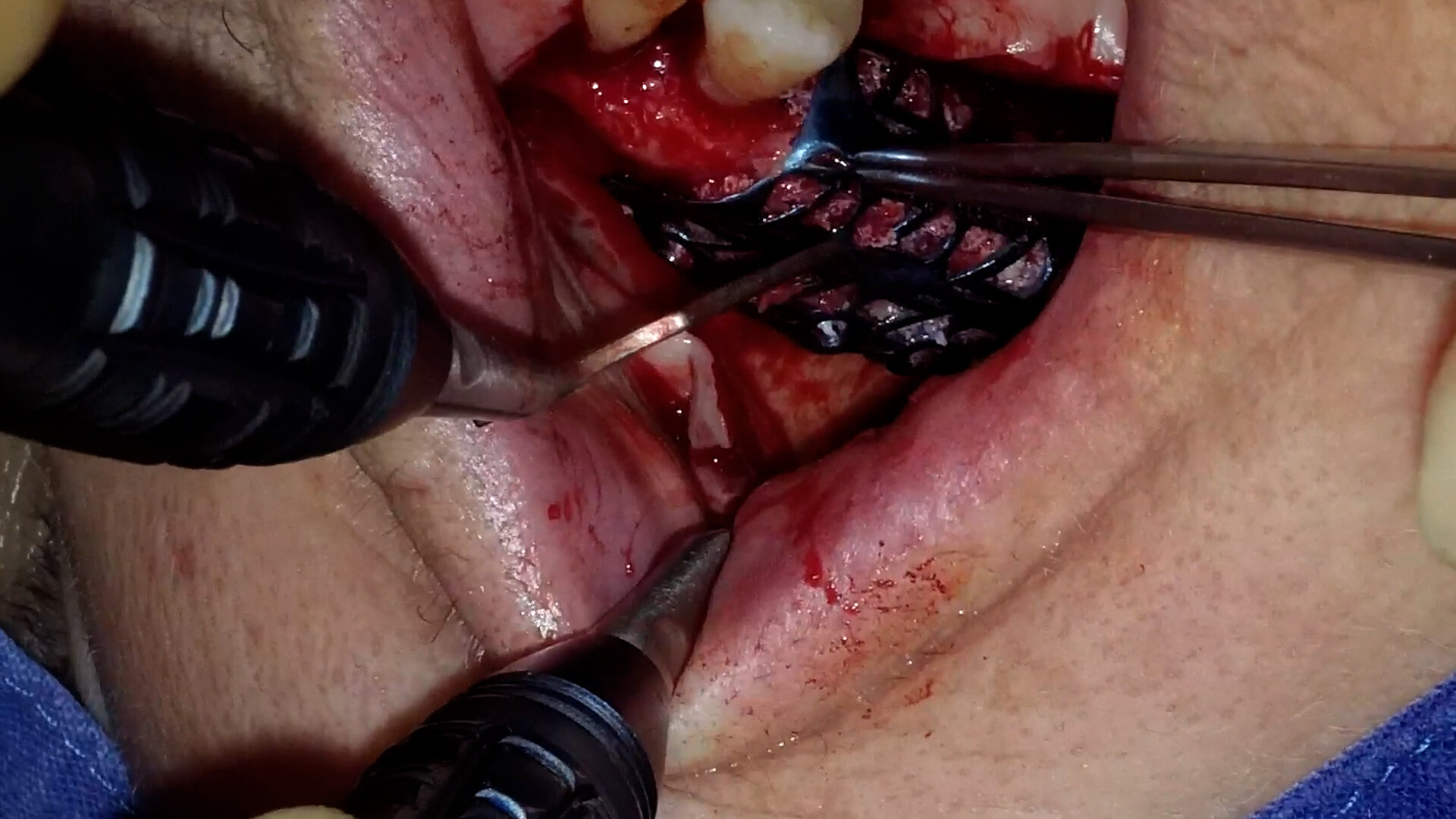
- The papilla preservation technique was performed using microsurgical instruments
- The root surface area was conditioned with 24% EDTA for ca. 2 minutes
- Emdogain was applied to the root surface
- The defect was filled with the Emdogain/Bio-Oss® mixture
- The wound was closed with two mattress sutures one horizontal mattress suture to secure the graft in place, and a second modified vertical mattress suture to tightly close the papilla
- A 5-0 suture was used for the horizontal mattress suture, and a 6-0 monofilament was used for the vertical mattress suture
- Postoperative care entailed rinsing the wound twice daily for 4 weeks with 0.2% chlorhexidine and ibuprofen analgesia on the first few days after surgery
Contents
The patient's jaw displayed a generalized loss of clinical attachment and alveolar bone. His general history was unremarkable; the patient was a non-smoker. Microbiological tests showed large numbers of Actinobacillus actinomycetemcomitans and Porphyromonas gingivalis. The diagnosis was "generalized aggressive periodontitis". After four months of initial therapy consisting of antibiotic combination therapy (amoxicillin + metronidazole), intraoral radiographs showed a deep and wide intraosseous bone defect located mesial and palatal to tooth #13. To preserve this strategically important tooth we opted to perform regenerative therapy with Emdogain and Bio-Oss cancellous bone material. Ten months after regenerative periodontal therapy, the probing depth had decreased by 7 mm, and 5-6 mm of clinical attachment had been gained. At this time, the probing depth was 2-3 mm and intraoral radiographs showed near-complete filling of the osseous defect.


