Quintessence International, 6/2025
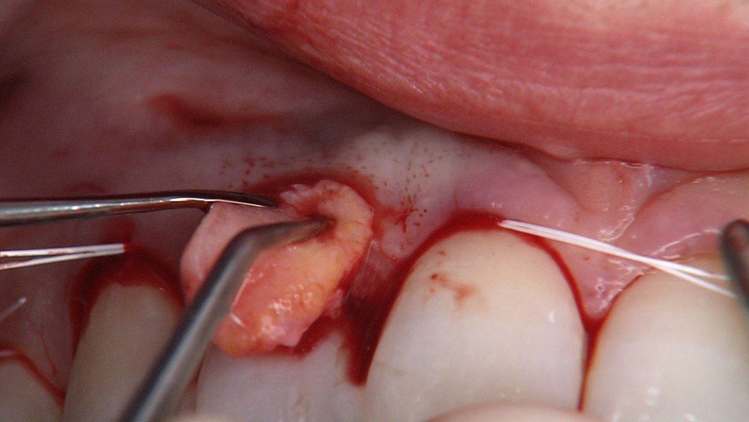
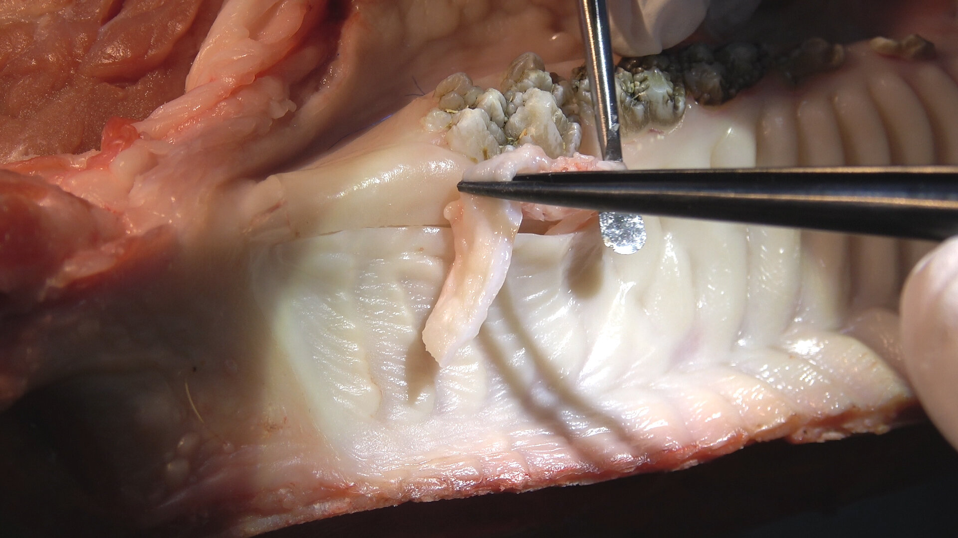
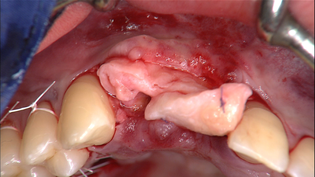
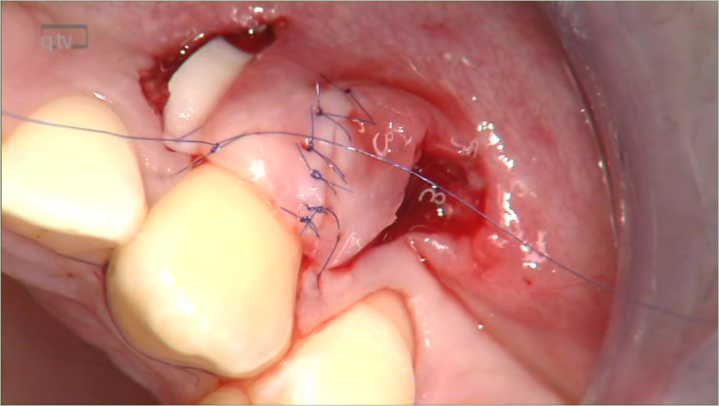
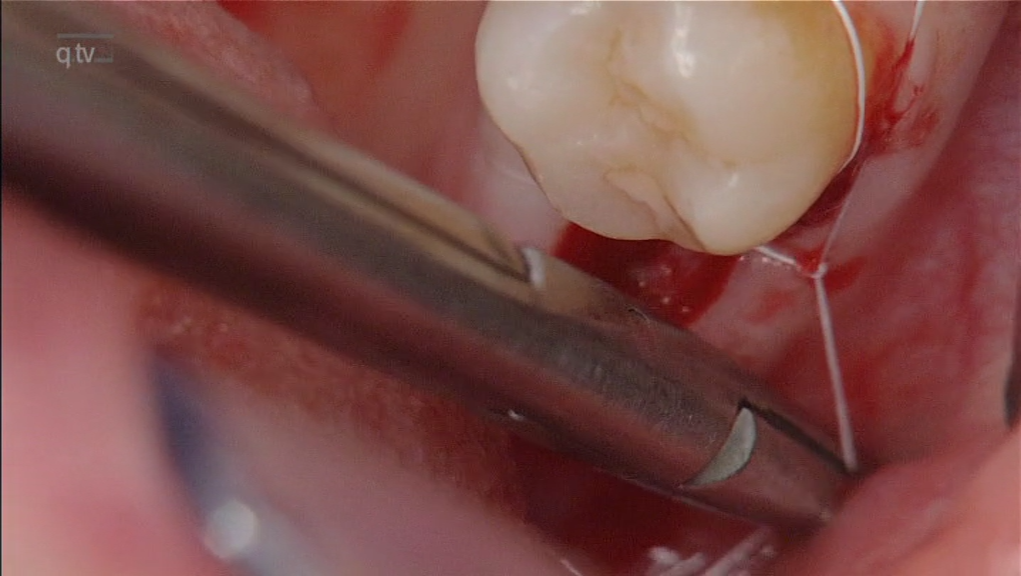
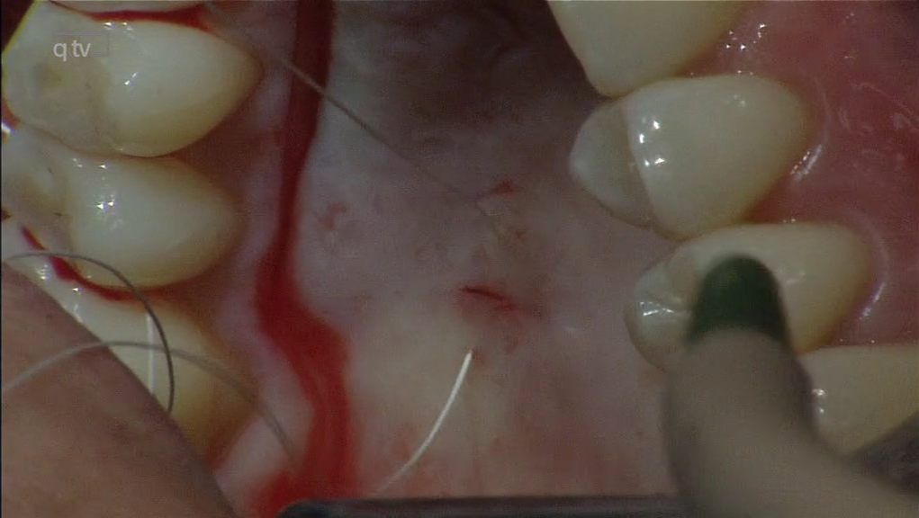
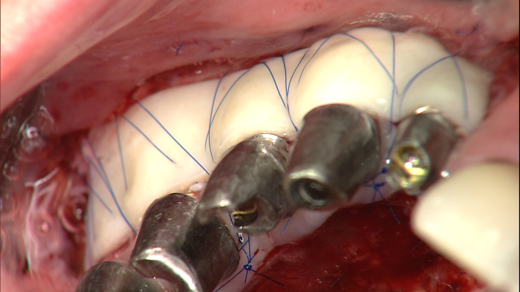
DOI: 10.3290/j.qi.b6245834, PubMed-ID: 40539426Seiten: 450-462, Sprache: EnglischGülnergiz, Erdem / Abraha, Sophia M. / Hürzeler, Markus / Zuhr, OttoNowadays, complication-free wound healing processes are the key to successful treatment outcomes in the context of periodontal and implant surgery, both clinically and scientifically. The main challenge here is to achieve primary wound healing in the majority of cases. Among the scientifically documented factors that influence the healing process, it is primarily the blood supply in the surgical area and the stability of the wound achieved postoperatively that can be directly influenced by the clinician. The surgical wound closure plays a decisive role in this context in order to achieve sufficient stabilization of the wound without negatively affecting the healing process through unnecessary traumatization of the tissue or excessive tensile forces on the wound edges. It is important to bear in mind that wound healing after surgical procedures in the oral cavity does not take place under optimal conditions. A moist, microbiologically contaminated environment is present and complete immobilization of the wound is hardly possible during the early healing phases. The sutures must therefore ensure that the surgical flaps are passively secured in the intraoperatively established position, that the wound edges are in as close contact as possible – especially if grafts that initially rely on nutrition through plasmatic circulation are used – and that the wound is stabilized during the first few postoperative days. The suture material and suturing technique must be selected so that the knots do not loosen and both the suture material and soft tissue can withstand the mechanical stresses during the early wound healing phases. The search for available mechanical anchors should be the focus of interest.
periodontal and implant surgery, suture anchors, suture materials, suture techniques
The International Journal of Oral & Maxillofacial Implants, 4/2025
DOI: 10.11607/jomi.11308Seiten: 459-467a, Sprache: EnglischLiao, Hung-Chi / Kan, Joseph Y.K. / Rungcharassaeng, Kitichai / Lin, Guo-Hao / Chen, Joey / Zuhr, Otto / Hürzeler, Markus / Lozada, JaimePurpose: To evaluate implant success rates and facial mucosal profile changes in maxillary single immediate implant placement and provisionalization with the socket-shield (IIPP+SS) technique and without the socket-shield (IIPP-SS) technique. Materials and Methods: A total of 30 dental implants in 25 patients were assigned to either the IIPP-SS group (15 implants) or the IIPP+SS (15 implants) group. Clinical and radiographic outcomes were collected preoperatively (T0) as well as at 2-week (T1), 6-month (T6), and 12-month (T12) postoperative follow-ups. The implant success rate, marginal bone level changes, facial mucosal level changes, and papilla level changes were evaluated at different time points. Facial mucosal profile changes were assessed individually for hard and soft tissue zones and as a whole using volumetric analysis. Results: Two implants were excluded (one patient dropped out and one implant failed) from the data analysis in this study, resulting in an overall implant success rate of 96.6% (28/29) after 1 year. Fewer facial mucosal profile changes were noted in the IIPP+SS group than in the IIPP-SS group; however, the difference was only marginally statistically significant (P = .06). No statistically significant difference was found in the facial mucosal level changes (P = .18) and papilla level changes (P = .67 for the mesial papilla level, P = .41 for the distal papilla level) between the IIPP-SS and IIPP+SS groups. Conclusions: Within the limitations of this 1-year randomized controlled clinical trial, the IIPP+SS group appeared to maintain the implant facial mucosal profile slightly better than IIPP alone. Both treatment modalities provide clinically satisfactory outcomes biologically, functionally, and esthetically.
Schlagwörter: dental implants, facial mucosal profile and level, immediate tooth replacement, implant esthetics, socket-shield technique
International Journal of Esthetic Dentistry (EN), 3/2025
Basic ResearchSeiten: 300-311, Sprache: EnglischGülnergiz, Erdem / Zuhr, Otto / Abraha, Sophia M. / Toll, Maximilian / Bastos, Joel / Hürzeler, MarkusA novel 3D classificationThe socket shield technique (SST) has gained great attention in the 14 years since its first publication. Different modifications of the original publication have been published in the meantime, with each one trying to become more predictable in the clinical outcome and prevent possible complications. For a better understanding of the mechanical and biologic principles of the SST, a novel 3D classification is developed and described in this article. This classification aims to help clinicians better anticipate and prevent the main long-term biologic complication of the SST, namely the coronal migration of the shield over time. In addition, this novel and clinically relevant 3D classification should allow for more effective and accurate communication between clinicians as well as a more objective assessment when it comes to the surgical execution of the SST.
Schlagwörter: immediate implants, implants in the esthetic zone, partial extraction therapy, socket shield technique, socket shield technique complications
Parodontologie, 2/2025
Seiten: 117-131, Sprache: DeutschGülnergiz, Erdem / Abraha, Sophia M. / Hürzeler, Markus / Zuhr, OttoEin UpdateHeute stehen im Zusammenhang mit parodontal- und implantatchirurgischen Eingriffen komplikationslose Wundheilungsabläufe als Schlüssel zu erfolgreichen Behandlungsresultaten klinisch wie auch wissenschaftlich im Mittelpunkt des Interesses. Die zentrale Herausforderung ist hierbei, in der Mehrheit der Fälle eine primäre Wundheilung zu erzielen. Unter den wissenschaftlich dokumentierten, den Heilungsprozess beeinflussenden Faktoren sind es vor allem die Blutversorgung im Operationsbereich sowie die postoperativ erzielte Stabilität der Wunde, auf die seitens des Behandlers Einfluss genommen werden kann. Die chirurgische Naht spielt in diesem Zusammenhang eine entscheidende Rolle, um einerseits eine ausreichende Stabilisierung der Wunde zu erreichen, ohne andererseits den Heilungsverlauf durch eine unnötige Traumatisierung des Gewebes oder übermäßige Zugkräfte auf die Wundränder negativ zu beeinträchtigen. Dabei gilt es zu berücksichtigen, dass die Wundheilung nach operativen Eingriffen in der Mundhöhle unter nicht optimalen Bedingungen stattfindet. Es liegt ein feuchtes, mikrobiologisch kontaminiertes Milieu vor und eine komplette Ruhigstellung der Wunde ist während der frühen Heilungsphasen kaum möglich. Die Nähte müssen deshalb für eine passive Sicherung der chirurgischen Lappen in der intraoperativ etablierten Position, einen möglichst innigen Kontakt der Wundränder sowie eine Stabilisierung der Wunde während der ersten postoperativen Tage sorgen. Die Suche nach verfügbaren mechanischen Ankerpunkten steht hierbei im Vordergrund des Interesses.
Schlagwörter: Parodontal- und Implantatchirurgie, Nahttechniken, Nahtanker, Nahtmaterialien
Quintessenz Zahnmedizin, 1/2022
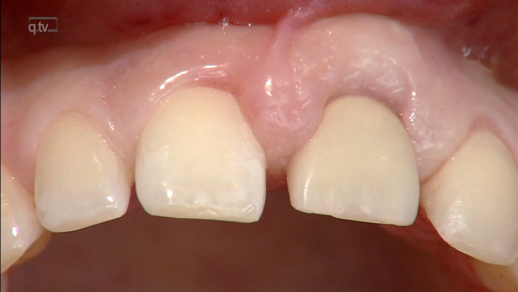
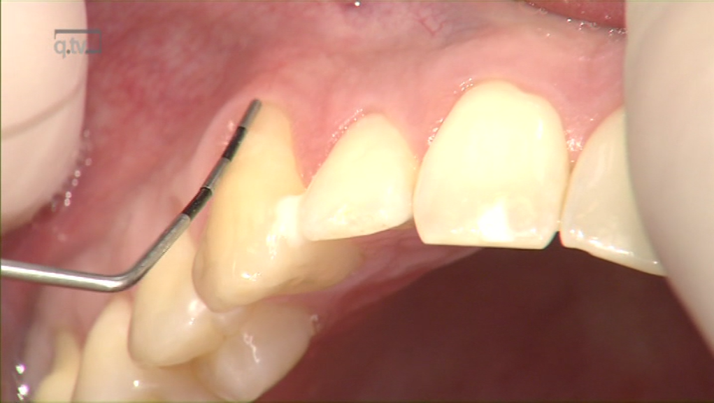
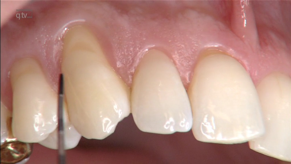
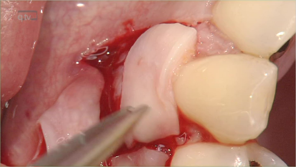
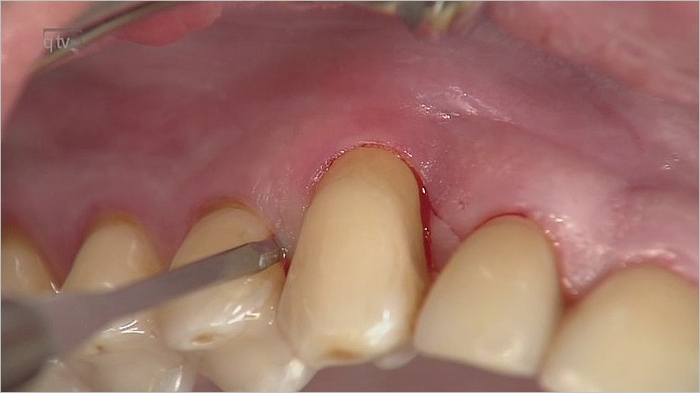
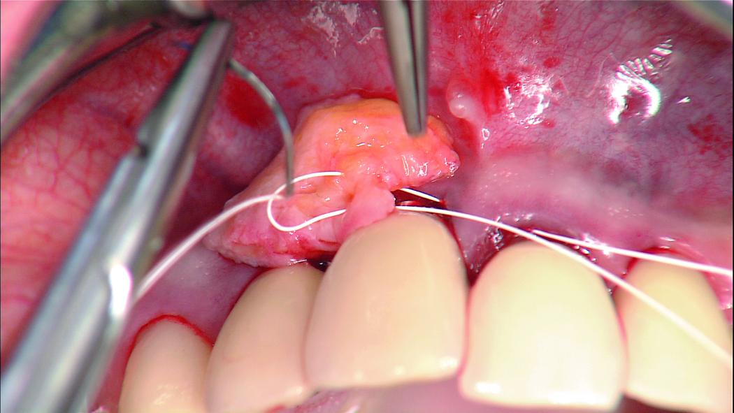
ProthetikSeiten: 52-57, Sprache: DeutschStähler, Philip / Gülnergiz, Erdem / Abraha, Sophia M / Zuhr, Otto / Hürzeler, MarkusAm vorliegenden Fall einer Adhäsivbrücke am lateralen Schneidezahn wird die ästhetisch motivierte Kieferkammaugmentation vorgestellt. Der heutzutage übliche weichgewebige Kieferkammaufbau erfolgt mit tunnelierenden Techniken sowie intraoral gewonnenen Bindegewebetransplantaten. Der gleichzeitige bindegewebige Kammaufbau mit Einbringen der prothetisch bereits ideal ausgeformten Adhäsivbrückenbasis ermöglicht die einzeitige Etablierung eines ästhetischen Emergenzprofils am Brückenglied.
Manuskripteingang: 07.10.2021, Annahme: 08.11.2021
Schlagwörter: Ästhetik, Weichgewebemanagement, Transplantat, Adhäsivbrücke
International Journal of Periodontics & Restorative Dentistry, 3/2020
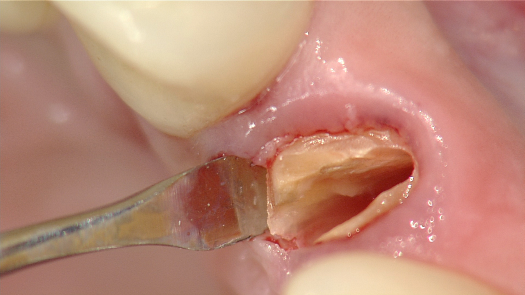
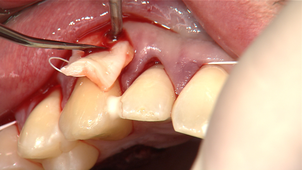
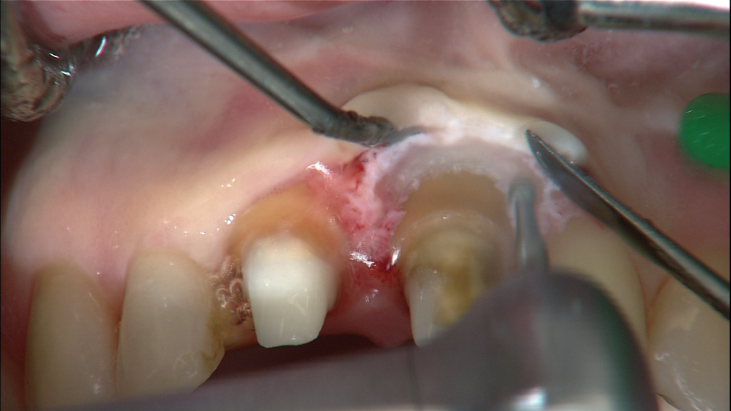
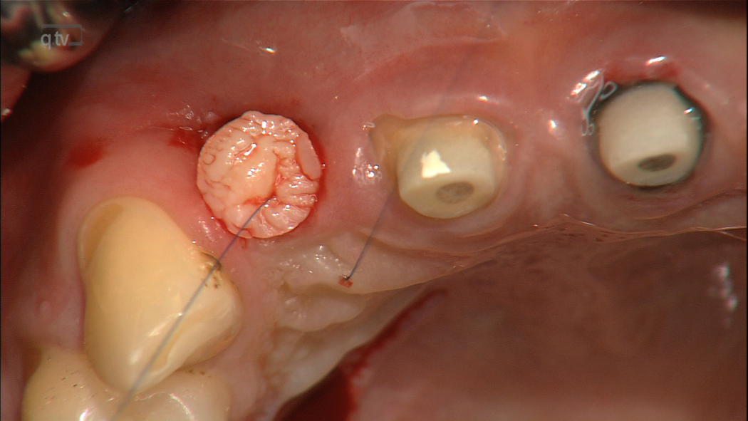
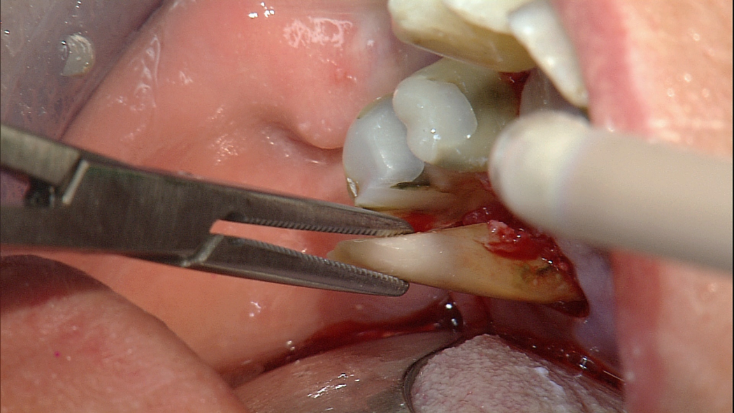
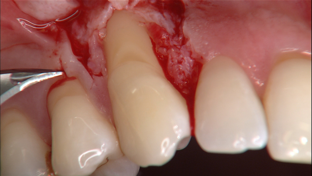
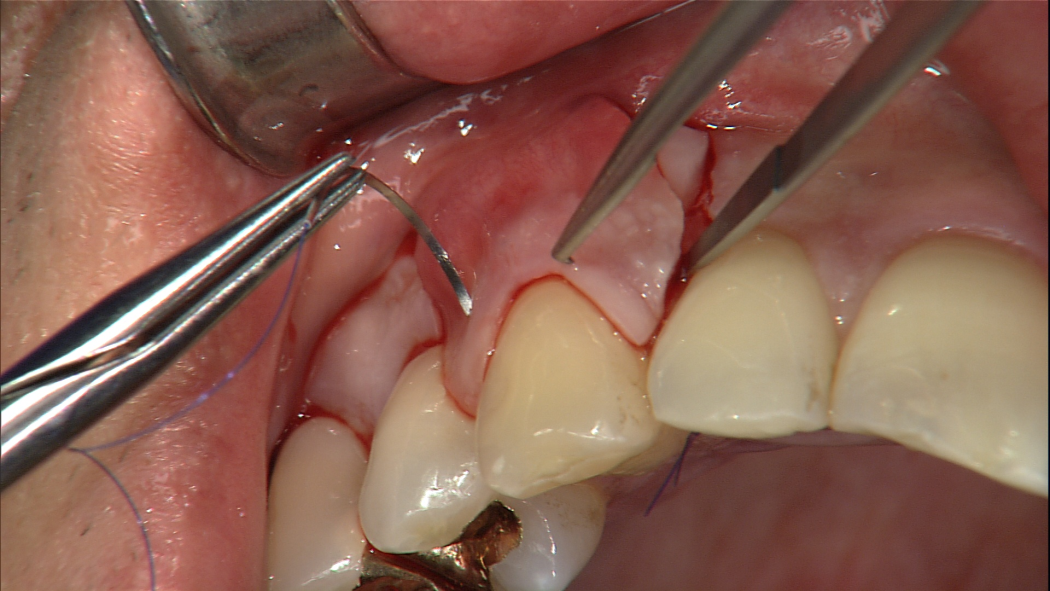
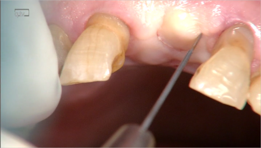
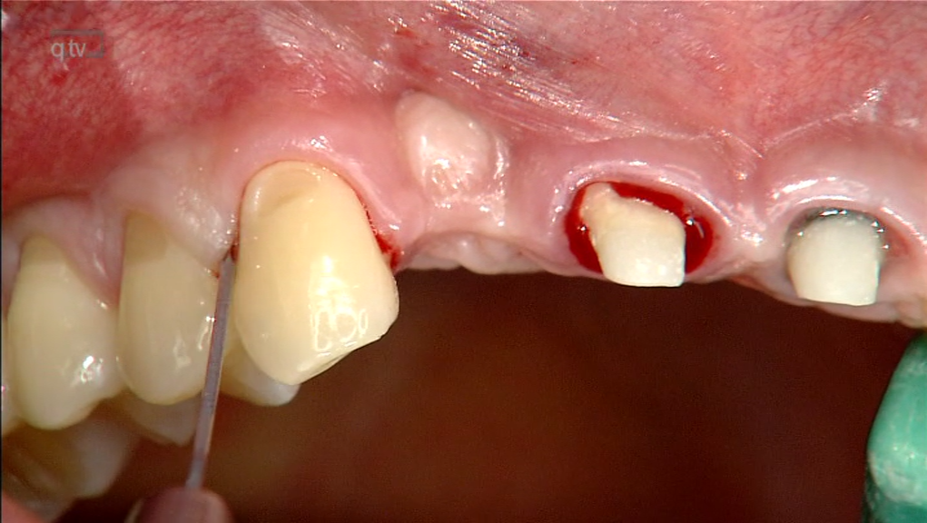
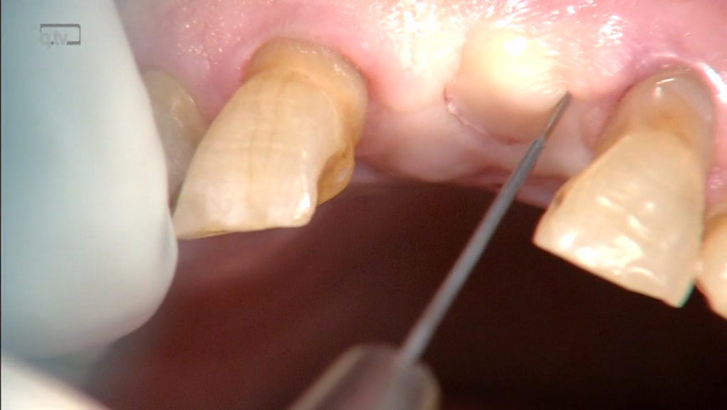
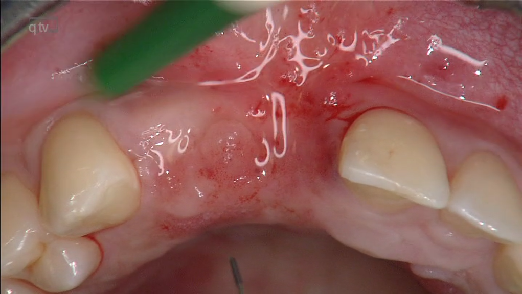
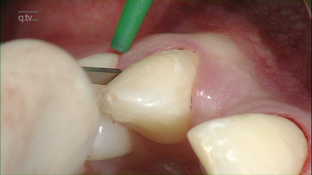
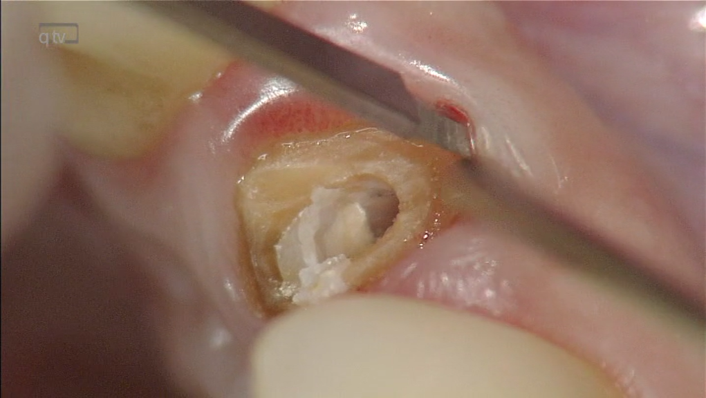
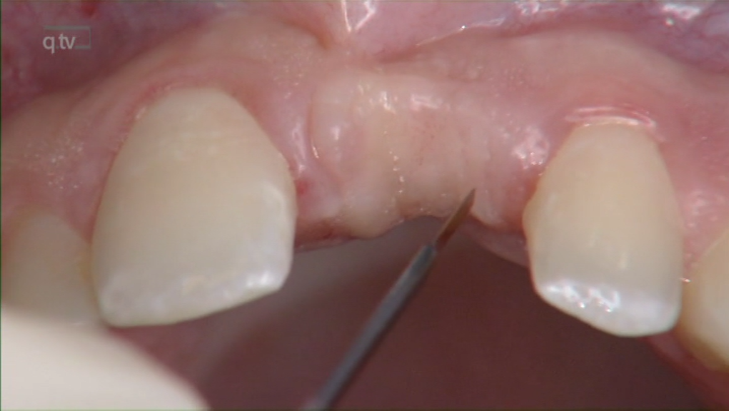
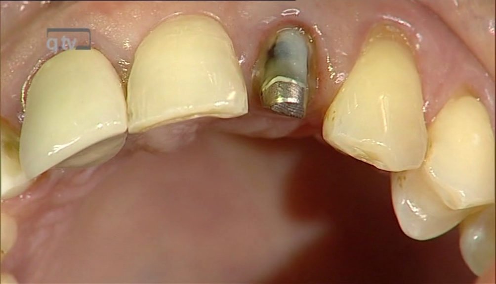
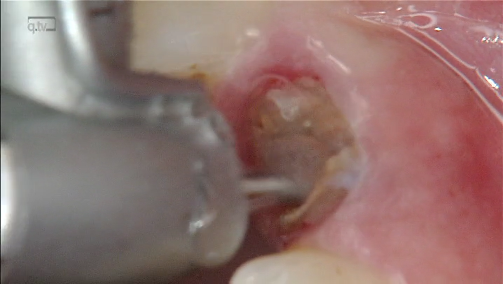
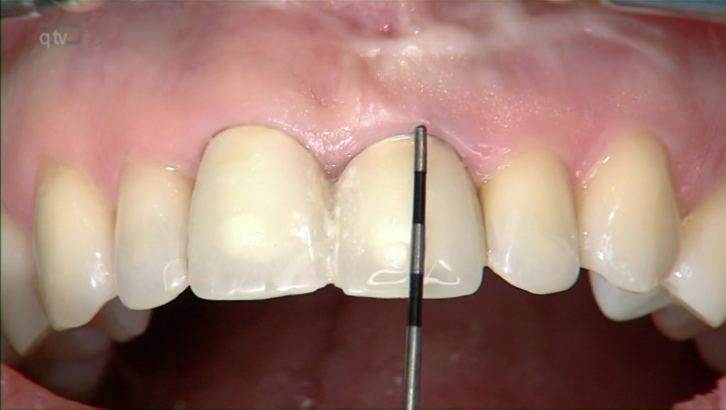
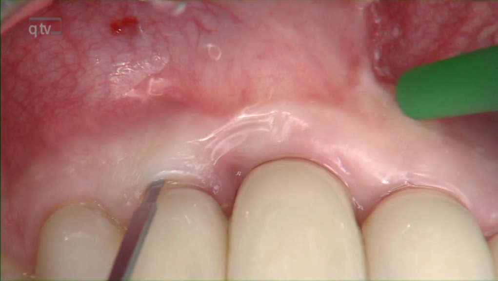
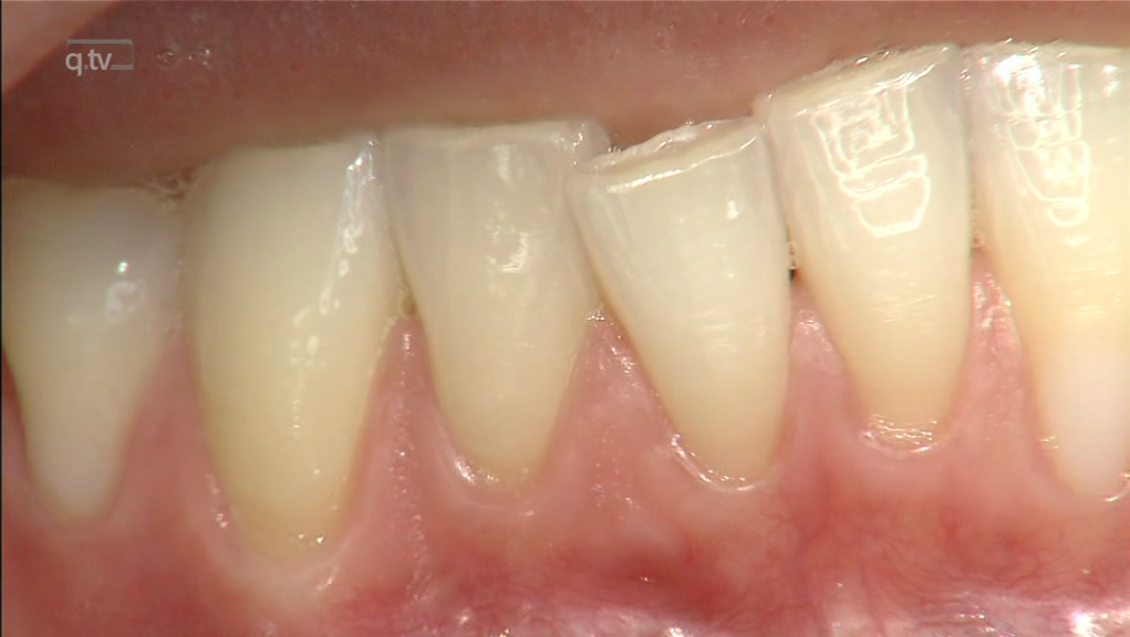
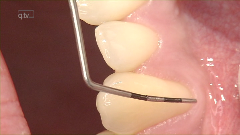
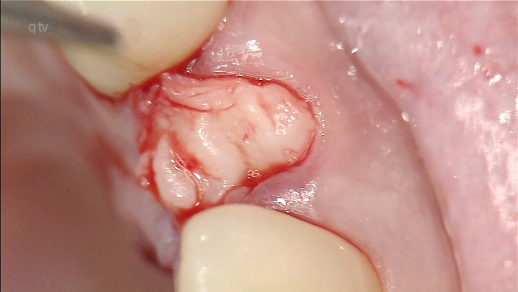
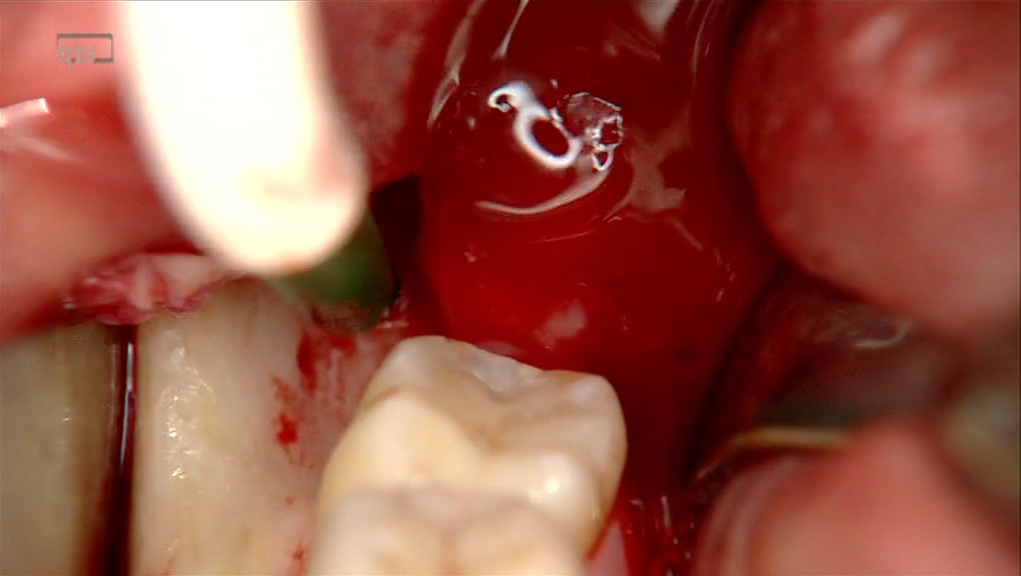
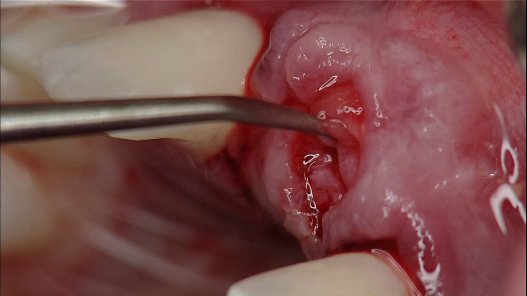
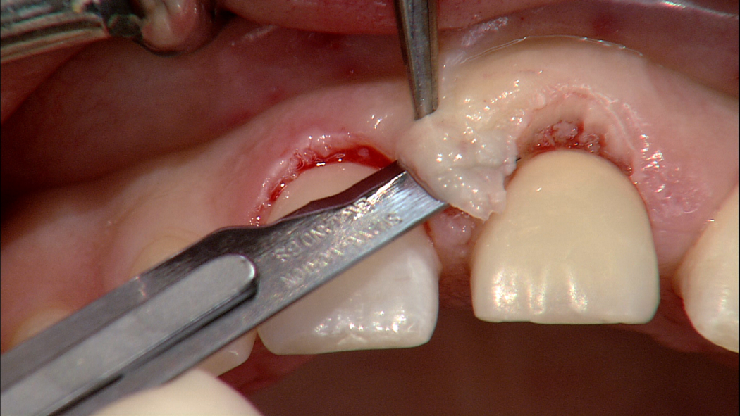
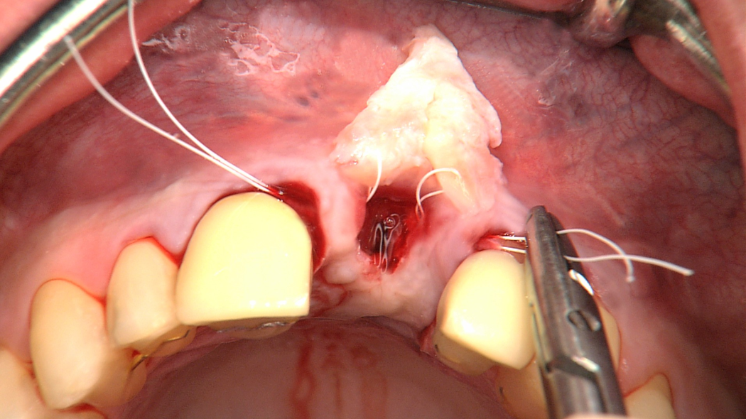
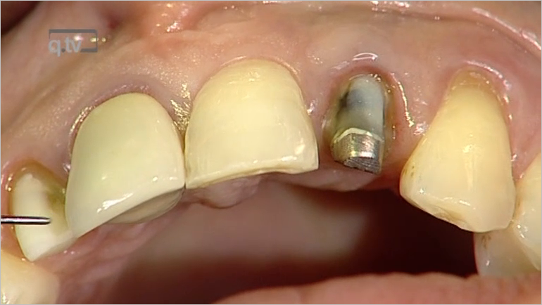
DOI: 10.11607/prd.4648, PubMed-ID: 32233195Seiten: 409-415, Sprache: EnglischZuhr, Otto / Staehler, Philip / Huerzeler, MarkusMaintaining soft and hard tissues around dental implants after tooth extraction is one of the major challenges in implant dentistry. After tooth extraction, the subsequent loss of bone and soft tissue is inevitable due to the partial resorption of the buccal bone plate. The recently described socket shield technique addresses the problem by maintaining the buccal piece of the tooth in the extraction socket in order to preserve the buccal bone. As with every new technique, specific complications, like infection of the buccal piece of the tooth, can occur. Herein, the authors present a clinical case that developed a complication with the socket shield technique and the consequential surgical management.
International Journal of Esthetic Dentistry (DE), 3/2020
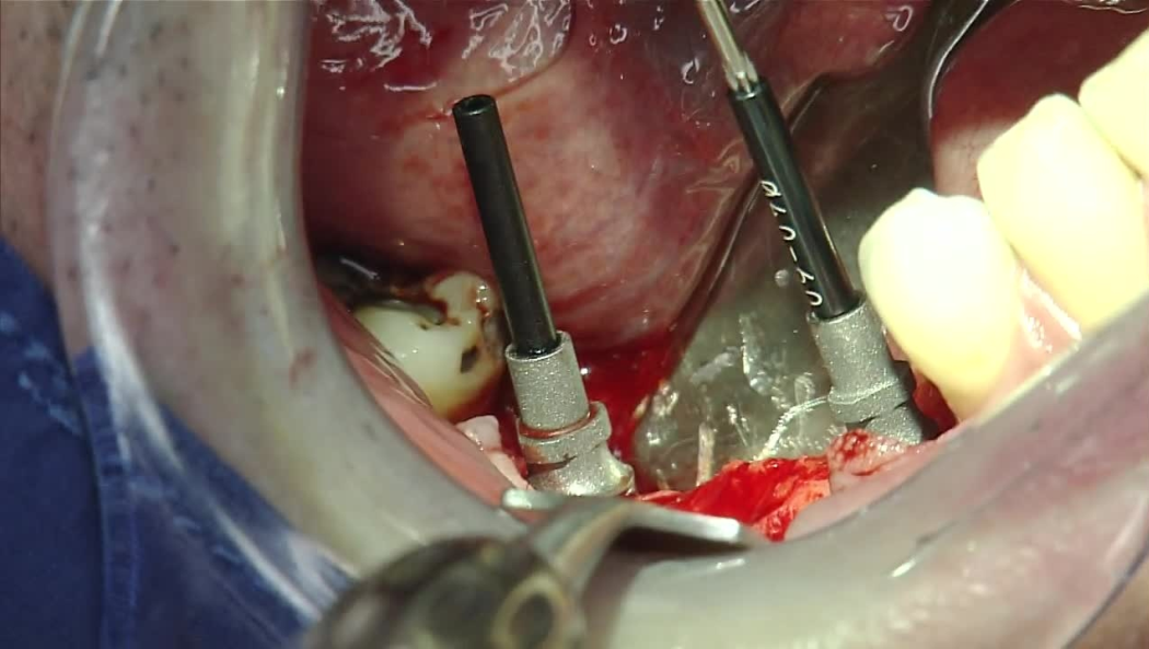
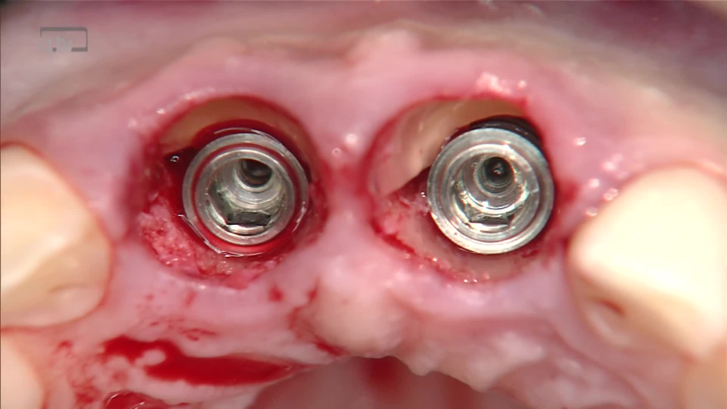
Seiten: 302-319, Sprache: DeutschStähler, Philip / Abraha, Sophia M. / Bastos, Joel / Zuhr, Otto / Hürzeler, MarkusDie Socket-Shield-Technik wurde erstmals im Jahr 2010 publiziert und ist inzwischen weltweit wissenschaftlich und klinisch anerkannt. Um möglichen Komplikationen bei diesem innovativen Ansatz für die ästhetische Implantologie zu begegnen, wird im vorliegenden Artikel ein umfassendes Schritt-für- Schritt-Protokoll vorgestellt, das die im letzten Jahrzehnt gemachten Erfahrungen mit aufnimmt. Nach der Dekoronation des Zahns wird das Implantatbett durch die Wurzel des nicht erhaltungswürdigen Zahns hindurch präpariert. Nach der Extraktion des palatinalen Wurzelfragments wird dann der Schild nach dem Prinzip einer mechanischen oder einer biologischen "Sperrung" präpariert. Die mechanische "Sperrung" wird durch direkten Kontakt des Implantats mit dem Schild bewirkt. Beim biologischen Ansatz hingegen wird versucht, eine Ankylose des Schildes herbeizuführen. Ziel ist, die Koronalverlagerung des Schildes zu verhindern. Der koronale Anteil des Schildes wird in eine konkave Form gebracht und endet 0,5 mm koronal des vestibulären Knochens. Anschließend wird das Implantat gesetzt und ein individueller Gingivaformer hergestellt. Wird die Socket-Shield-Technik im Einklang mit den zugrunde liegenden biologischen und mechanischen Prinzipien ausgeführt, liefert sie vorhersagbar hochästhetische Resultate.
International Journal of Esthetic Dentistry (EN), 3/2020
PubMed-ID: 32760924Seiten: 288-305, Sprache: EnglischStaehler, Philip / Abraha, Sophia M. / Bastos, Joel / Zuhr, Otto / Hürzeler, MarkusThe socket-shield technique, first published in 2010, has gained worldwide scientific and clinical acceptance. To address possible complications with this innovative approach in esthetic implant dentistry, we provide a comprehensive step-by-step protocol incorporating what we have learnt in the past decade. After initial decoronation of the tooth, the implant bed is prepared through the root of the tooth to be extracted. Following extraction of the palatal root fragments, the shield is prepared according to either a mechanical or biologic 'locking' principle. The mechanical 'locking' comprises a direct contact between the implant and the shield, whereas the biologic approach facilitates ankylosis of the shield, preventing its coronal displacement. The coronal part of the shield is brought into a concave shape, ending up 0.5 mm coronal to the buccal bone. The implant is consequently inserted, and an individualized healing cap fabricated. When performed according to the underlying biologic and mechanical principles, the socket-shield technique can provide highly esthetic and predictable outcomes.
Kieferorthopädie, 1/2020
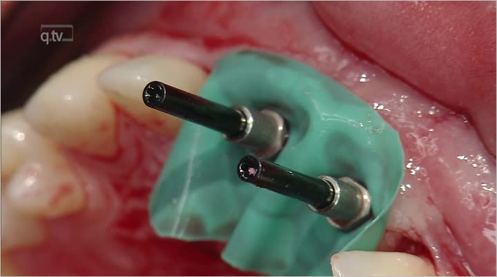
Interdisziplinäre KieferorthopädieSeiten: 47-51, Sprache: DeutschHürzeler, Markus / Zuhr, Otto / Richter, Wolf / Ludwig, Björn / Zimmermann, Moritz / Hürzeler, Bärbel / Schoberer, UliDie Behandlung von Erwachsenen nimmt einen hohen Anteil im kieferorthopädischen Alltag ein. Erwachsenenbehandlungen zeichnen sich in der Kieferorthopädie meist durch eine hohe Komplexität aus und erfordern daher das Zusammenspiel mehrerer Fachdisziplinen, um das Behandlungsziel zu erreichen. In der neuen Rubrik "Interdisziplinäre Kieferorthopädie" werden komplexe interdisziplinäre Patientenbeispiele vorgestellt. Der Beitrag basiert auf der Originalpublikation Hürzeler et al. Int J Esthet Dent 2019;14:116–117.
Schlagwörter: ästhetische Zone, interdisziplinäre Kieferorthopädie, Miniimplantate, vertikale Augmentation, implant site development
International Journal of Esthetic Dentistry (DE), 2/2019
Seiten: 124-125, Sprache: DeutschHürzeler, Markus / Zuhr, Otto / Richter, Wolf / ludwig, Björn / Hürzeler, Bärbel / Schoberer, Uli