Wir verwenden Cookies ausschließlich zu dem Zweck, technisch notwendige Funktionen wie das Login oder einen Warenkorb zu ermöglichen, oder Ihre Bestätigung zu speichern. Mehr Informationen zur Datenerhebung und -verarbeitung finden Sie in unserer Datenschutzerklärung.
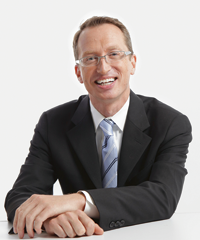
Dr. Blatz ist Professor für restaurative Zahnmedizin, Vorsitzender der Abteilung für präventive und restaurative Wissenschaften und stellvertretender Dekan für digitale Innovation und berufliche Weiterbildung an der University of Pennsylvania School of Dental Medicine in Philadelphia, wo er das Penn Dental Medicine CAD/CAM Ceramic Center gründete. Er ist Redaktionsmitglied von Quintessence International und einer der Autoren des internationalen Bestsellers Evolution: Aktuelle Konzepte für Einzelzahnimplantate im Frontzahnbereich (Quintessence Publishing, 2014). Dr. Blatz wurde mehrfach ausgezeichnet und ist ein gefragter Referent zu den Themen ästhetische Zahnmedizin, restaurative Materialien und Implantologie.
Voraussichtliches Erscheinen: Juni 2025 1. Auflage 2025 Buch Softcover; 13.5 x 19 cm, 220 Seiten, 220 Abbildungen Sprache: Englisch Kategorien: Zahnheilkunde allgemein, Ästhetische Zahnheilkunde, Prothetik ISBN 978-1-78698-142-4 QP Deutschland
Aktuelle Konzepte für Einzelzahnimplantate im Frontzahnbereich
1. Auflage 2016 Buch Hardcover, 27,8 x 27, 8 cm, 440 Seiten, 1985 Abbildungen Sprache: Deutsch Kategorien: Implantologie, Literatur fürs Studium ISBN 978-3-86867-257-2 QP Deutschland
10. Okt. 2024 — 12. Okt. 2024Palacio de congresos de Sevilla, Sevilla, Spanien
Referenten: Sam Alawie, Markus B. Blatz, Marco Ferrari, Henriette Terezia Lerner, Antonio Liñares, Arturo Llobell, Ignazio Loi, Michel Magne, Pascal Magne, Francesco Mangano, Eitan Mijiritsky, Nazariy Mykhaylyuk, Mutlu Özcan, George Papavasiliou, Alessandro Pozzi, Marta Revilla-León, Vygandas Rutkūnas, Teresa Sierpińska, Jacobo Somoza, Miguel Stanley, Francesca Vailati
SEPES - Sociedad Española de Prótesis Estomatologías y Estética
35th EAED Spring Open Meeting
35 years of Esthetic Dentistry - The masters, the fundamentals, today's trends23. Mai 2024 — 25. Mai 2024The Baron's Hall & Gallery, Vila Nova Gaia, Portugal
Referenten: Luís Azevedo, Urs C. Belser, Nitzan Bichacho, Markus B. Blatz, Jorge Cardoso, Nuno Sousa Dias, Petra Gierthmühlen, Aiste Gintaute, Oscar Gonzalez-Martin, Stefano Gracis, Ueli Grunder, Arndt Happe, Marc Hürzeler, France Lambert, Amélie Mainjot, Alexandros Manolakis, Konrad H. Meyenberg, Nazariy Mykhaylyuk, José M. Navarro, Gaetano Paolone, Stavros Pelekanos, Roberto Perasso, João Pitta, Pablo Ramírez, Andrea Ricci, Giano Ricci, Giuseppe Romeo, Irena Sailer, David Winkler, Yu Zhang, Giovanni Zucchelli
European Academy of Esthetic Dentistry
The 9th World Dental Meeting in Japan 2023
No Dentistry, No Wellness!29. Sept. 2023 — 1. Okt. 2023Pacifico Yokohama Conference Center, Yokohama, Japan
Referenten: Alessandro Agnini, Andrea Mastrorosa Agnini, Wael Att, Gustavo Avila-Ortiz, Markus B. Blatz, Victor Clavijo, Karim Dada, Glécio Vaz de Campos, Vincent Fehmer, Naoki Hayashi, Mario Imburgia, Guillaume Jouanny, Sérgio Kahn, Bertrand Khayat, Sandra Khong Tai, Christopher Köttgen, Stefen Koubi, Tomas Linkevičius, Nazariy Mykhaylyuk, Ravindra Nanda, Andreas Nolte, Léon Parienté, Jose Manuel Reuss , Domenico Ricucci, Isabella Rocchietta, Irena Sailer, Todd R. Schoenbaum, Werner Schupp, Istvan Urban, Eric Van Dooren, Débora R. Vilaboa, Otto Zuhr
Quintessence Publishing Co. Ltd. Japan
ISC 2023 – The 26th International Symposium on Ceramics
Simplicity Meets Esthetics8. Juni 2023 — 11. Juni 2023Town and Country Resort, San Diego, Vereinigte Staaten von Amerika
Referenten: Wael Att, Diego Bechelli, Markus B. Blatz, Domenico Cascione, Gerard J. Chiche, Stephen J. Chu, Victor Clavijo, Florin Cofar, Iván Contreras Molina, Don Cornell, Lee Culp, Vincent Fehmer, Mirela Feraru, German O. Gallucci, Petra Gierthmühlen, Gustavo Giordani, David Guichet, Galip Gürel, Effrat Habsha, Hilal Kuday, Kenneth A. Malament, Adam J. Mieleszko, Ricardo Mitrani, Masayuki Okawa, Jacinthe M. Paquette, Joshua Polansky, Marta Revilla-León, Giuseppe Romeo, Irena Sailer, John Sorensen, Martina Stefanini, Taiseer Sulaiman, Douglas A. Terry, Van Thompson, George Tysowsky, Eric Van Dooren, Yu Zhang
Referenten: Nitzan Bichacho, Markus B. Blatz, Christian Coachman, Luca De Stavola, Mirela Feraru, Gustavo Giordani, Galip Gürel, Ronald Jung, Nazariy Mykhaylyuk, Nelson R. F. A. Silva, Dennis P. Tarnow
SEPES - Sociedad Española de Prótesis Estomatologías y Estética
The 14th International Symposium on Periodontics and Restorative Dentistry (ISPRD)
9. Juni 2022 — 12. Juni 2022Boston Marriott Copley Place, Boston, MA, Vereinigte Staaten von Amerika
Referenten: Tara Aghaloo, Edward P. Allen, Evanthia Anadioti, Wael Att, Vinay Bhide, Markus B. Blatz, Scotty Bolding, Lorenzo Breschi, Jeff Brucia, Daniel Buser, Luigi Canullo, Daniele Cardaropoli, Stephen J. Chu, Donald Clem, Christian Coachman, Lyndon F. Cooper, Daniel Cullum, Lee Culp, José Carlos Martins da Rosa, Sergio De Paoli, Marco Degidi, Nicholas Dello Russo, Serge Dibart, Joseph P. Fiorellini, Mauro Fradeani, Stuart J. Froum, David Garber, Maria L. Geisinger, William Giannobile, Luca Gobbato, Ueli Grunder, Galip Gürel, Chad Gwaltney, Christoph Hämmerle, Robert A. Horowitz, Marc Hürzeler, David Kim, Gregg Kinzer, Christopher Köttgen, Ina Köttgen, Purnima S. Kumar, Burton Langer, Lydia Legg, Pascal Magne, Kenneth A. Malament, Jay Malmquist, George Mandelaris, Pamela K. McClain, Michael K. McGuire, Mauro Merli, Konrad H. Meyenberg, Craig M. Misch, Julie A. Mitchell, Marc L. Nevins, Myron Nevins, Michael G. Newman, Miguel A. Ortiz, Jacinthe M. Paquette, Stefano Parma-Benfenati, Michael A. Pikos, Giulio Rasperini, Pamela S. Ray, Christopher R. Richardson, Isabella Rocchietta, Marisa Roncati, Marco Ronda, Paul S. Rosen, Maria Emanuel Ryan, Irena Sailer, Maurice Salama, David M. Sarver, Takeshi Sasaki, Todd Scheyer, Massimo Simion, Michael Sonick, Sergio Spinato, Dennis P. Tarnow, Lorenzo Tavelli, Douglas A. Terry, Tiziano Testori, Carlo Tinti, Istvan Urban, Hom-Lay Wang, Robert Winter, Giovanni Zucchelli
Quintessence Publishing Co., Inc. USA
Zeitschriftenbeiträge dieses Autors
International Journal of Periodontics & Restorative Dentistry, Pre-Print
DOI: 10.11607/prd.7277, PubMed-ID: 3943672722. Okt. 2024,Seiten: 1-22, Sprache: EnglischShakibaie, Behnam / Nava, Paolo / Calatrava, Javier / Blatz, Markus B. / Nagy, Katalin / Sabri, Hamoun
This prospective, preliminary controlled clinical trial investigates the comparative effectiveness of platform-switching (PS) versus traditional butt-joint or platform-matching (PM) implant-abutment connections on peri-implant crestal bone stability. Utilizing a split mouth design, 10 systemically healthy patients (n= 20 implants) had adjacent non-restorable maxillary anterior teeth replaced with two different implants (butt-joint connections and platform-switching interfaces). Patients underwent alveolar ridge preservation, followed by implant placement: platform-matching implants were inserted at crestal bone level, and platform-switching implants were placed 1mm subcrestally. Customized Zirconia crowns were then fabricated for both systems. Outcome measures included bleeding on probing (BOP), probing pocket depth (PPD), and marginal bone loss (MBL), which were evaluated through standardized periapical radiographs over 3-year timeframe. Results showed significantly higher initial MBL in the PM group (0.86 ± 0.13 mm) compared to the PS group (0.34±0.29 mm) [p value: p<0.01]. Moreover, at the three-year follow-up, the crestal bone levels remained above the implant shoulder until the third year of the study for the PS subcrestal group (PS: -0.15±0.39 mm) and slightly below the implant platform in the PM crestal group (PM: 0.55±0.19). After 3 years, the PS group also exhibited lower mean BOP percentages (12%) than the butt-joint group (17%). This study suggests that subcrestal placement with PS and internal connections can provide better long-term peri- implant bone preservation, thereby potentially improving implant success and aesthetic outcomes in the anterior maxillary region.
Schlagwörter: dental implant, abutment, dental implant-abutment connection, platform-switching, platform-matching, implant-supported dental prostheses, marginal bone levels
The International Journal of Prosthodontics, Pre-Print
DOI: 10.11607/ijp.8880, PubMed-ID: 391109302. Aug. 2024,Seiten: 1-21, Sprache: EnglischRivera, Macarena / Angulo, Alexandra / Cerda, Barbara / Schulz, Rolando / Blatz, Markus B.
Purpose: To evaluate the preferences of dental practitioners for tooth-supported crowns dental ceramics by means of an online survey and to assess the influence of age, gender, years of experience, and dental specialty on those preferences. Materials and Methods: An anonymous questionnaire was delivered online through Google Surveys, targeting 796 dentists. It contained 17 dichotomous, closed questions with two sections. The first section dealt with general characteristics, including age, gender, years of experience, training in prosthodontics, and hours worked per week. The second section included questions regarding preferences of different materials for crowns, and the use of digital workflows. Analyses were carried out with Stata 14.0 software (StataCorp, TX, USA). A significance level of p=0.05 was adopted. Results: 248 surveys were answered. Practitioners in Chile preferred lithium disilicate for the anterior region (55.2%) but also for the posterior area (40.7%), regardless of their age, gender, years since graduation, hours worked per week or any training in prosthodontics. Chilean dentists over 50 years old considered zirconia almost 4 times more than those under 30 years old for anterior crowns (51.85%). 59.68% of dentists take digital impressions, and 37.10% have access to chairside CAD/CAM technology. In this group, 54.4% preferred feldspathic ceramic for anterior and 23.9% for posterior crowns. Conclusions: While there is a wide range of dental ceramic materials, these results provide a snapshot of the current trends in Chile where lithium disilicate is the most preferred ceramic material for tooth-supported crowns, and metal-ceramic is the least preferred material.
Schlagwörter: Ceramics, survey, zirconia, CAD/CAM, questionnaire
Ziel: Ziel dieser retrospektiven Fallserie war eine Längsschnittuntersuchung der Volumenveränderungen des vestibulären Weichgewebevolumens an Implantaten bezogen auf die Dicke und die Breite der keratinisierten Mukosa (DKM bzw. BKM) nach Weichgewebeaugmentation mittels Bindegewebetransplantat (BGT) und mikrochirurgischer Envelope-Technik.
Material und Methoden: Bei 12 gesunden Patienten wurden 12 Implantate im Ober- oder Unterkieferseitenzahnbereich eingesetzt. Die Studie umfasste die minimalinvasive Entnahme von 12 BGT mit einer Einzelinzisionstechnik und deren Transplantation zur Augmentation des vestibulären periimplantären Weichgewebes unter Verwendung einer Envelope-Technik. Anschließend wurden die Implantate mit 12 verschraubten Keramikkronen versorgt (IPS e.max).
Ergebnisse: Die Heilung verlief an allen Implantaten unauffällig und die Patienten wurden anschließend 5 Jahre nachbeobachtet. Für die DKM war in den ersten 6 postoperativen Wochen der größte Rückgang zu beobachten (von 5,50 ± 0,79 mm auf 4,59 ± 0,62 mm); anschließend sank sie nochmals leicht ab (auf 4,00 ± 0,85 mm) und blieb bis zur 2-Jahres-Nachuntersuchung stabil (bei 4,00 ± 0,36 mm). Zwischen dem zweiten und dem dritten Jahr nach der Operation nahm die DKM nochmals ab (auf 3,59 ± 0,42 mm) und blieb dann bis zum Ende des 5-jährigen Beobachtungszeitraums konstant. Die Beobachtungen zur BKM waren hiervon leicht verschieden: Die Messungen zeigten, dass die größte Abnahme in den ersten 6 Wochen stattfand (von 2,50 ± 0,42 auf 1,50 ± 0,42 mm), und die BKM anschließend bis zur 1-Jahres-Nachuntersuchung gehalten wurde. Vom ersten zum zweiten Jahr nach dem Eingriff stieg die BKM wieder an (auf 2,00 ± 0,60 mm) und blieb während der nächsten 3 Jahre gleich (bei 2,00 ± 0,85 mm).
Schlussfolgerungen: Die vorliegende Untersuchung konnte die Vorteile deutlich machen, welche die Kombination aus periimplantärer Weichgewebeaugmentation mittels eines minimalinvasiv entnommenen BGT und der mikrochirurgischen Envelope-Technik über einen Zeitraum von 5 Jahren bietet.
Schlagwörter: Bindegewebetransplantat, Envelope-Technik, Implantologie, Mikrochirurgie, periimplantäres Weichgewebe
Aim: The aim of the present retrospective case series was to longitudinally assess soft tissue volume changes on the vestibular aspect of implants in relation to keratinized mucosa thickness (KMT) and width (KMW) after the application of the microsurgical envelope technique combined with a connective tissue graft (CTG).
Materials and methods: A total of 12 healthy patients received 12 dental implants placed either in the posterior maxilla or mandible. The study involved the harvesting of 12 CTGs with a minimally invasive single-incision technique, grafted to the vestibular peri-implant soft tissue utilizing the envelope technique, followed by the insertion of 12 screw-retained IPS e.max crowns.
Results: The healing process was uneventful across all areas, and all patients were followed up for a period of 5 years. The evaluation of KMT showed the highest decrease in the first 6 weeks after surgery (5.5 ± 0.79 to 4.59 ± 0.62 mm), then dropped slightly to 4 ± 0.85 mm, after which it maintained at 4 ± 0.36 mm until the 2-year time point. Between the second and third years after surgery, a further decrease of 3.59 ± 0.42 mm was recorded for KMT, which then remained constant until the end of the 5-year research period. The observations regarding KMW were slightly different, with the measurements demonstrating the greatest decrease in first 6 weeks (from 2.5 ± 0.42 to 1.5 ± 0.42 mm), which was maintained until the 1-year time point. Between the first and second years after surgery, the KMW increased to 2 ± 0.60 mm and remained level for the next 3 years, at 2 ± 0.85 mm.
Conclusions: The current research demonstrated the advantages of using a combination of a minimally invasively harvested CTG and the microsurgical envelope technique for a duration of 5 years.
Schlagwörter: connective tissue graft, envelope technique, implantology, microsurgery, peri-implant soft tissue
International Journal of Computerized Dentistry, 1/2023
ApplicationDOI: 10.3290/j.ijcd.b3818287, PubMed-ID: 36825568Seiten: 75-88, Sprache: Englisch, DeutschConejo, Julián / Isleem, Wael / Atria, Pablo J. / Ayub, José María / Baumgarten, Harold S. / Blatz, Markus B.
Die Digitalisierung der Zahnmedizin hat während der letzten zehn Jahre viel zur Entwicklung und Vereinfachung der zahnärztlichen Implantologie beigetragen. Dank Intraoralscannern, digitaler Volumentomografie, 3-D-Implantatplanungssoftware und CAD/CAM-Systemen ist die prothetisch orientierte Implantologie zu einem unproblematischen Verfahren geworden. Digitale Hilfsmittel für die Planung und Durchführung von Behandlungen nach evidenzbasierten Konzepten verbessern die langfristige Ästhetik und Funktion implantatgetragener Restaurationen ebenso wie das Langzeitüberleben von Dentalimplantaten. Letztere gelten häufig als erste Behandlungsoption für den Ersatz nicht erhaltungswürdiger oder fehlender Zähne. Implantatversorgungen in der ästhetischen Zone bleiben jedoch für viele Zahnärztinnen und Zahnärzte eine Herausforderung. Dieser Artikel liefert klinische Leitlinien für eine ideale Implantatpositionierung mittels computergeführter Chirurgie sowie für chairside CAD/CAM-gefertigte provisorische und definitive Implantatkronen mit Titanbasen, die gute prothetische Ergebnisse, eine optimale Gingivaarchitektur und eine Verkürzung der Behandlungsdauer ermöglichen.
Schlagwörter: Dentalimplantat, Implantatrestauration, chairside, CAD/CAM, Titanbasis, Implantatposition, Sofortprovisorium, schablonengeführte Implantatchirurgie
Background and aim: Dental implant patients are frequently required to undergo a second-stage/uncovery procedure to expose the implant fixture. The aim of the present prospective study was to evaluate the clinical outcomes of the vestibular split rolling flap (VSRF) versus the double door mucoperiosteal flap (DDMF) techniques at adjacent posterior implant sites during the second-stage procedure.
Materials and methods: A total of 44 uncovered posterior dental implants in 10 healthy patients were treated at the second stage. All the mesial implants were assigned to the VSRF technique (group A) and the distal implants to the DDMF technique (group B). Soft tissue measurements were performed as vestibular keratinized mucosal width (KMW) and vestibular mucosal thickness (MT) over a period of 1 year, assessed at four different intervals.
Results: Healing was uneventful at all sites. There were no patient dropouts in the entire study time frame. The clinical comparison of the adjacent implants showed overall higher MT measurements at 12 months for group A (2.5 ± 0.2 mm) compared with group B (1.00 ± 0.3 mm), and for KMW measurements for group A (2.5 ± 0.2 mm) compared with group B (2.0 ± 0.3 mm).
Conclusions: The VSRF technique described in the present article is a reliable method for performing an implant uncovery. If the technique is applied according to the indication and with a minimally invasive protocol, it is preferable to other conventional exposure techniques due to its ability to provide enhanced soft tissue volume around the implant, which can in turn benefit the health, esthetics, function, and long-term stability of the peri-implant tissue.
Hintergrund und Ziel: In vielen Fällen ist nach einer Implantation ein zweiter Eingriff zur Freilegung der Implantatfixtur notwendig. Ziel der vorliegenden prospektiven Studie war ein Vergleich der klinischen Ergebnisse bei Anwendung folgender Freilegungstechniken an benachbarten Seitenzahnimplantaten: vestibulärer Teilschicht-Rolllappen (vestibular split rolling flap, VSRF) und Doppeltür-Mukoperiostlappen (double door mucoperiosteal flap, DDMF).
Material und Methoden: Insgesamt 44 gedeckt eingeheilte Implantate bei 10 gesunden Patienten wurden bei einem Zweiteingriff freigelegt. Die jeweils mesialen Implantate wurden der VSRF-Technik (Gruppe A), die distalen der DDMF-Technik (Gruppe B) zugeteilt. Zur Bewertung des Weichgewebes wurden während eines Jahres an vier Zeitpunkten die Breite der vestibulären keratinisierten Mukosa (KMB) und die Dicke der vestibulären Mukosa (MD) gemessen.
Ergebnisse: Die Heilung verlief an allen Implantaten unauffällig. Während der gesamten Studiendauer gab es keine Studienabbrüche. Im klinischen Vergleich der benachbarten Implantate ergaben sich nach 12 Monaten höhere MD-Werte in Gruppe A (2,5 ± 0,2 mm) als in Gruppe B (1,00 ± 0,3 mm) und höhere KMB-Werte in Gruppe A (2,5 ± 0,2 mm) als in Gruppe B (2,0 ± 0,3 mm).
Schlussfolgerungen: Die im vorliegenden Artikel beschriebene VSRF-Technik ist ein zuverlässiges chirurgisches Verfahren zur Implantatfreilegung. Wird diese Technik indikationsgemäß und nach einem minimalinvasiven Protokoll angewendet, liefert sie im Gegensatz zu anderen konventionellen Freilegungstechniken ein größeres Weichgewebevolumen um Implantate. Dies wirkt sich günstig auf die Gesundheit, Ästhetik, Funktion und langfristige Stabilität des periimplantären Gewebes aus.
Implant position and soft tissue thickness have a direct influence on implant abutment design. The goal is to place the implant in the optimal spatial position to maintain the adjacent bone and soft tissues. When the implant is not placed ideally, prosthetic variations to abutments and restorations must be made, which may limit the esthetic appearance of the final restoration or alter the biologic environment of the bone and tissues. This article illustrates and explains the effect of different implant positions on the emergence profile design in order to assist the clinician with treatment planning and selection in various clinical situations.
New digital tools facilitating data acquisition, team communication, computer-assisted diagnostics, and treatment planning as well as the design and fabrication of restorations, guides, stents and devices in general have fundamentally altered key clinical and laboratory steps. The number of new technologies and the amount of new equipment used today to acquire patient data, the software to manipulate this data, and the machines to manufacture devices from it drastically increases all the time, as do the challenges of integrating these systems into a feasible, realistic, and practical workflow. Creating a simple complete digital workflow is key to taking advantage of these digital opportunities and offering their benefits to all patients. Making digital workflows the routine rather than the exception is fundamentally important in order to grow a dental practice in this new environment. This article presents a new complete digital workflow that changes and improves the process of treating a comprehensive case from diagnostics to execution and maintenance.
Neue digitale Möglichkeiten für die Datenakquise, Kommunikation im Team, computergestützte Diagnostik und Behandlungsplanung sowie Design und Herstellung von Restaurationen, Schablonen, Schienen und Apparaturen im Allgemeinen haben zu grundlegenden Veränderungen bei den zentralen klinischen und zahntechnischen Arbeitsschritten geführt. Täglich erhöht sich die Anzahl neuer Techniken und Geräte, die mittlerweile für die Erhebung von Patientendaten eingesetzt werden. Es gibt stetig neue Software für die Verarbeitung dieser Daten und es entstehen neue Geräte, die anhand dieser Daten Restaurationen und Apparaturen herstellen. Damit wachsen jedoch auch die Herausforderungen, diese Systeme in sinnvolle, realistische und praktikable Arbeitsabläufe einzubinden. Die Etablierung eines einfachen volldigitalen Workflows ist der Schlüssel dazu, die digitalen Möglichkeiten sinnvoll und zum Vorteil der Patienten einzusetzen. Um eine Zahnarztpraxis vor diesem neuen Hintergrund erfolgreich zu führen, müssen digitale Workflows von der Ausnahme zur Regel gemacht werden. In diesem Artikel wird ein neuer volldigitaler Workflow vorgestellt, um das Management komplexer Fälle von der Diagnostik über die Behandlung bis zur Nachsorge zu verändern und verbessern.