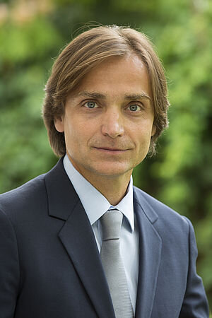
Wir verwenden Cookies ausschließlich zu dem Zweck, technisch notwendige Funktionen wie das Login oder einen Warenkorb zu ermöglichen, oder Ihre Bestätigung zu speichern. Mehr Informationen zur Datenerhebung und -verarbeitung finden Sie in unserer Datenschutzerklärung.
Dr. Urban erhielt seinen DMD-Abschluss und anschließend seinen MD-Abschluss an der Semmelweis University School of Medicine and Dentistry (Budapest, Ungarn) in den Jahren 1991 und 1996. Er absolvierte ein Vollzeitstudium der Oralchirurgie am St. Istvan Hospital in Budapest, Ungarn (1992-1996). Sein Praktikum in Parodontologie absolvierte er an der UCLA. Nach Abschluss des Fellowship-Programms (1999-2000) in Implantologie an der Loma Linda University in Loma Linda, Kalifornien, wurde er im folgenden Jahr zum Assistenzprofessor ernannt. Dr. Urban unterrichtet Implantologie im Rahmen des Graduiertenprogramms der Loma Linda University. Er ist im Staat Kalifornien (USA) zugelassen und hat eine Privatpraxis in Budapest, Ungarn. Dr. Urban promovierte 2012 in Parodontologie an der Universität von Szeged, Ungarn. Derzeit ist er Honorarprofessor an der Universität von Szeged. Dr. Urban ist Vorstandsmitglied der Osteology Foundation und hat wissenschaftliche Artikel und Fachbücher über Knochenregeneration und rekonstruktive Weichgewebschirurgie bei Zahnimplantaten veröffentlicht.
1. Auflage 2022 Buch Hardcover, 21 x 28 cm, 560 Seiten, 2050 Abbildungen Sprache: Englisch Kategorien: Implantologie, Mund-Kiefer-Gesichtschirurgie Artikelnr.: 7701 ISBN 978-1-78698-108-0 QP Deutschland
1. Auflage 2018 Buch Hardcover, 21 x 28 cm, 400 Seiten, 1252 Abbildungen Sprache: Deutsch Kategorie: Implantologie ISBN 978-3-86867-372-2 QP Deutschland
1st Edition 2017 Buch Hardcover, 21 x 28 cm, 400 Seiten, 1252 Abbildungen Sprache: Englisch Kategorie: Implantologie Artikelnr.: 7479 ISBN 978-1-78698-000-7 QP Deutschland
Bone and Soft Tissue Regeneration22. März 2024 — 23. März 2024São Paulo, Brasilien
Referenten: Fouad Khoury, José Carlos Martins da Rosa, Istvan Urban, Giovanni Zucchelli
The 9th World Dental Meeting in Japan 2023
No Dentistry, No Wellness!29. Sept. 2023 — 1. Okt. 2023Pacifico Yokohama Conference Center, Yokohama, Japan
Referenten: Alessandro Agnini, Andrea Mastrorosa Agnini, Wael Att, Gustavo Avila-Ortiz, Markus Blatz, Victor Clavijo, Karim Dada, Glécio Vaz de Campos, Vincent Fehmer, Naoki Hayashi, Mario Imburgia, Guillaume Jouanny, Sérgio Kahn, Bertrand Khayat, Christopher Köttgen, Stefen Koubi, Tomas Linkevičius, Nazariy Mykhaylyuk, Ravindra Nanda, Andreas Nolte, Léon Parienté, Jose Manuel Reuss , Domenico Ricucci, Isabella Rocchietta, Irena Sailer, Todd R. Schoenbaum, Werner Schupp, Sandra Tai, Istvan Urban, Eric Van Dooren, Débora R. Vilaboa, Otto Zuhr
Quintessence Publishing Co. Ltd. Japan
Pikos • Urban
5. Mai 2023 — 6. Mai 2023The Ritz-Carlton Orlando, Grande Lakes, Orlando, Vereinigte Staaten von Amerika
Referenten: Michael A. Pikos, Istvan Urban
Pikos Institute
International Osteology Symposium
27. Apr. 2023 — 29. Apr. 2023Barcelona, Spanien
Osteology Foundation
4th Buser & Sculean International Symposium
Regeneration and Esthetics in Periodontology and Implant Dentistry2. Dez. 2022 — 3. Dez. 2022Congress Center, Kursaal Bern, Bern, Schweiz
Referenten: Sofia Aroca, Daniel Buser, Stephen Chen, Massimo De Sanctis, Nikolaos Donos, Markus B. Hürzeler, Sascha Jovanovic, Ronald Jung, Giulio Rasperini, Isabella Rocchietta, Frank Schwarz, Anton Sculean, Shakeel Shahdad, Andreas Stavropoulos, Martina Stefanini, Leonardo Trombelli, Istvan Urban, Giovanni Zucchelli
Buser & Sculean Academy
3rd Urban International Hard and Soft Tissue Regeneration Symposium
Predictability in Regeneration21. Okt. 2022 — 22. Okt. 2022Budapest Congress Center, Budapest, Ungarn
Referenten: Luca De Stavola, Ueli Grunder, Jaime Lozada, José Carlos Martins da Rosa, Aušra Ramanauskaitė, Frank Schwarz, Istvan Urban, Giovanni Zucchelli, Otto Zuhr
Urban Regeneration Institute
29th EAO annual scientific meeting
Uniting Nations through Innovations29. Sept. 2022 — 1. Okt. 2022Palexpo, Le Grand-Saconnex, Schweiz
Referenten: Thabo Beeler, Nitzan Bichacho, Rino Burkhardt, Luigi Canullo, Matteo Chiapasco, Luca De Stavola, Mirela Feraru, Alfonso Gil, Klaus Gotfredsen, Markus Gross, Ueli Grunder, Christoph Hämmerle, Björn Klinge, Ivo Krejci, Sebastian Kühl, Niklaus P. Lang, Sonia Leziy, Daniele Manfredini, Konrad H. Meyenberg, Francesco Mintrone, Ricardo Mitrani, Sven Mühlemann, José M. Navarro, Florian Probst, Pablo Ramírez, Christoph Andreas Ramseier, Mario roccuzzo, Fidel Ruggia, Mariano Sanz, Rubens Spin-Neto, Dennis P. Tarnow, Daniel Thoma, Istvan Urban, Hans-Peter Weber
European Association for Osseintegration (EAO)
EuroPerio10
15. Juni 2022 — 18. Juni 2022Bella Center Copenhagen, Copenhagen, Dänemark
Referenten: Mario Aimetti, Zvi Artzi, Serhat Aslan, Georgios Belibasakis, Florian Beuer, Juan Blanco Carrión, Michael M. Bornstein, Nagihan Bostanci, Philippe Bouchard, Darko Božić, Olivier Carcuac, Maria Clotilde Carra, Nelson Carranza, Iain L. C. Chapple, Pierpaolo Cortellini, Jan Cosyn, Mike Curtis, Francesco D'Aiuto, Bettina Dannewitz, Massimo De Sanctis, Luca De Stavola, Jan Derks, Nikolaos Donos, Peter Eickholz, Bahar Eren Kuru, Ricardo Faria Almeida, Roberto Farina, Magda Feres, Elena Figuero, Dagmar Fosså Bunæs, Rok Gašperšič, William Giannobile, Cecilie Gjerde Gjengedal, Moshe Goldstein, Marjolaine Gosset, Klaus Gotfredsen, Filippo Graziani, Adrian Guerrero, George Hajishengallis, Hady Haririan, Lisa J. A. Heitz-Mayfield, Palle Holmstrup, Markus B. Hürzeler, Mark Ide, Søren Jepsen, Ronald Jung, Sérgio Kahn, Anhgela R. Kamer, Alpdogan Kantarci, Moritz Kebschull, Björn Klinge, Thomas Kocher, Odd Carsten Koldsland, Kenneth Kornman, Purnima Kumar, Marja Laine, Markus Laky, Isabelle Laleman, Evanthia Lalla, France Lambert, Luca Landi, Niklaus P. Lang, Antonio Liñares, Tomas Linkevičius, Bruno Loos, Rodrigo Lopez, Eli Machtei, Aslan Mammadov, Mauro Merli, Andrea Mombelli, Eduardo Montero, Niki Moutsopoulos, Jose Nart, Gustavo G. Nascimento, Ian Needleman, Tiernan O'Brien, William Papaioannou, Panos N. Papapanou, Michael A. Pikos, Pawel Plakwicz, Constanza Pontarolo, Philip M. Preshaw, Marc Quirynen, Mia Rakic, Christoph Andreas Ramseier, Hélène Rangé, Papageorgiou Spyridon, Maurizio S. Tonetti, Leonardo Trombelli, Istvan Urban, Fridus van der Weijden, Fabio Vignoletti, Charalambos Vlachopoulos, Nicola West, Asaf Wilensky, Ion Zabalegui, Egija Zaura, Nicola Zitzmann, Giovanni Zucchelli, Otto Zuhr, Fardal Øystein
European Federation of Periodontology (EFP)
Zeitschriftenbeiträge dieses Autors
International Journal of Periodontics & Restorative Dentistry, Pre-Print
DOI: 10.11607/prd.6667, PubMed-ID: 375521848. Aug. 2023,Seiten: 1-17, Sprache: EnglischUrban, Istvan A / Saleh, Muhammad H A / Serroni, Matteo / Shahbazi, Arvin / Baksa, Gabor / Szoke, Peter / Ravid? , Andrea
Vertical ridge augmentation in the anterior mandible is a technically delicate procedure that requires knowledge of some anatomical structures to reduce peri- and post-operative complications. Proper soft tissue management is one of the primary aspects to the success of these techniques, enabling tension-free primary wound closure and preventing membrane exposure. In this cadaveric and clinical study, we provided an anatomical overview of the lingual portion of the anterior mandible. Moreover, we described a novel surgical approach for the release of the lingual flap that will help clinicians achieve primary closure without incurring intrasurgical complications.
Implants with deficient papillae and black triangle are common findings. The treatment of these esthetic complications is considered challenging, and with limited predictability. Therefore, the aim of the present report is to describe a novel technique for papilla augmentation (the "Iceberg" connective tissue graft [iCTG]) after extraction and interproximal bone reconstruction in the anterior region. A 35-year-old patient presented with a hopeless tooth with interproximal clinical attachment loss extending up the apical third of the adjacent tooth. Interproximal bone reconstruction was performed through alveolar ridge preservation by directly applying recombinant human platelet-derived growth factor-BB (rhPDGF-BB) to the exposed root surface of the adjacent tooth. A mixture of autogenous bone chips (obtained from the ramus) and bovine bone xenograft particles, previously mixed with the growth factor, was also used. The patient was able to come back for implant therapy only 2 years later. An incomplete regeneration of the interproximal bone was observed. Therefore, to compensate the interproximal deficiency, the iCTG approach, involving a double layer CTG with different origins, was utilized. Two small grafts from the tuberosity were sutured to the mesial and distal ends of a wider CTG harvested from the palate, aiming at gaining additional volume at the interproximal sites. The composite graft was then sutured on top of the implant platform, with the flap that was then released and closed by primary intention. After conditioning of the peri-implant tissues, the case was finalized with a satisfactory outcome. The described iCTG could be an effective approach for reconstructing peri-implant papilla following interproximal bone reconstruction.
International Journal of Periodontics & Restorative Dentistry, 2/2024
DOI: 10.11607/prd.6458, PubMed-ID: 37722007Seiten: 213-218, Sprache: EnglischShahbazi, Arvin / Windisch, Péter / Tubbs, R. Shane / Decater, Tess / Urbán, István A. / Baksa, Gábor / Iwanaga, Joe
Guided bone regeneration (GBR) requires a tension-free flap without damaging the collateral circulation in order to secure better surgical outcomes. Topographic knowledge regarding the neurovascular bundles in the posterior aspect of the mandible can prevent complications during lingual flap design. The lingual branch (LB) of the inferior alveolar or maxillary arteries is not sufficiently illustrated or described in the literature. Nevertheless, it has an intimate relationship with the lingual nerve (LN) during ridge augmentation and implant-related posterior mandible surgery. Therefore, this study aimed to clarify the morphology and topography of the LB related to GBR surgeries. In the present human cadaveric study, the LB was analyzed in 12 hemimandibles using latex injection and corrosion casting. Two types of LB were identified based on their origin and course. The LB was found in a common connective tissue sheath close to the LN. The LB assembled several anastomoses on the posterior lingual aspect of the mandible and retromolar area. The LB acted as an anatomical landmark in identifying LN at the posterior lingual aspect of the mandible.
International Journal of Periodontics & Restorative Dentistry, 1/2024
DOI: 10.11607/prd.6667Seiten: 17-25, Sprache: EnglischUrban, Istvan A. / Saleh, Muhammad H. A. / Serroni, Matteo / Shahbazi, Arvin / Baksa, Gabor / Szoke, Peter / Ravid? , Andrea
Vertical ridge augmentation in the anterior mandible is a technically delicate procedure that requires knowledge of some anatomical structures to reduce intra- and postoperative complications. Proper soft tissue management is one of the primary aspects to the success of these techniques, enabling tension-free primary wound closure and preventing membrane exposure. This cadaveric and clinical study provides an anatomical overview of the lingual portion of the anterior mandible. Moreover, there is a description of a novel surgical approach for release of the lingual flap that will help clinicians achieve primary closure without incurring intrasurgical complications.
This pilot case series study reports the dimensional alveolar bone changes after reconstruction of severely resorbed postextraction sockets treated with a mixture of particulate bone allograft and xenograft in combination with titanium-reinforced dense polytetrafluoroethylene (Ti-d-PTFE) membranes. Ten subjects who required premolar or molar extraction were included. Bone grafts were protected with Ti-d-PTFE membranes, utilizing an open-healing environment; membranes were removed 4 to 6 weeks after extraction, and implants were placed 6.7 months (mean) after extraction (T1). One patient required additional augmentation to correct an apical undercut of the alveolar process that was present preextraction. All implants integrated well and showed an implant stability quotient (ISQ) value between 71 and 83. The mean horizontal ridge width reduction from baseline (extraction) to T1 was 0.8 mm. Throughout the study, the mean vertical bone gain increase ranged from 0.2 mm to 2.8 mm (mean keratinized tissue width increase: 5.8 mm). The ridge preservation/restoration technique showed good preservation and restoration of severely resorbed sockets as well as improved amounts of keratinized tissue. If implant therapy is required after tooth extraction and severely resorbed sockets are present, the use of a Ti-d-PTFE membrane is a realistic option.
Introduction: Papilla reformation is one of the most difficult and elusive surgical techniques for clinicians. Although it involves similar tenets to those applied for soft tissue grafting at recession defects, crafting a small tissue in restricted space remains unpredictable. Numerous grafting techniques have been developed to correct interproximal and buccal recession, but so far, only a limited number of techniques have been prescribed for interproximal remediation.
Case presentation: This report describes in detail a modern technique (the vertical interproximal tunnel approach) for reforming the interproximal papilla and treating interproximal recession. It also documents three challenging cases of papilla loss. The first case presented Class II papilla loss and a recession type 3 gingival defect adjacent to a dental implant, managed using the vertical interproximal tunnel approach through a short vertical incision. A 6-mm increase in attachment level and almost complete papilla fill were observed in this case with this surgical technique for papilla reconstruction. The second and third cases presented Class II papilla loss between two adjacent teeth, managed using the vertical interproximal tunnel approach through a semilunar incision and achieving full papilla reconstruction.
Conclusion: Both described incision designs for the vertical interproximal tunnel approach require technical meticulousness. When executed carefully and using the most beneficial pattern of blood supply, predictable reconstruction of the interproximal papilla can be achieved. It also helps alleviate concerns associated with inadequate flap thickness, blood supply and flap retraction.
Schlagwörter: complications, gingival recession, peri-implantitis, root coverage
The authors declare they have no conflicts of interest. They do not have any financial interests, either directly or indirectly, in the products or information listed in the paper.
Various surgical flap advancement techniques for bone regeneration have been described in the literature; however, the clinical challenges of managing tissue that contains scars or embedded foreign materials have not been thoroughly described, especially around metal foramen. Fibrotic and thickened scar periosteum as well as mental foramen restrict the tissue from responding in the same way as native tissue. Therefore, additional considerations and approaches must be considered to achieve tension-free flap closure. This article presents a flap advancement classification that describes three common clinical scenarios based on the periosteum and soft tissue quality and provides surgical approaches for tissue management in each classification, with a focus on flap advancement around the mental foramen.
International Journal of Periodontics & Restorative Dentistry, 5/2022
DOI: 10.11607/prd.6055Seiten: 577-585, Sprache: EnglischUrban, Istvan A / Tattan, Mustafa / Ravida, Andrea / Saleh, Muhammad Ha / Tavelli, Lorenzo / Avila-Ortiz, Gustavo
Severe alveolar ridge deficiencies in concomitance with periodontal attachment loss can represent a serious clinical challenge in the context of implant therapy. The present case report describes the management of a complex defect in the esthetic zone via ridge augmentation and periodontal regenerative therapy using a biologic material. A systemically healthy 55-year-old man diagnosed with peri-implantitis around an implant in the maxillary left central incisor position and with severe bone loss on the mesial aspect of the maxillary left lateral incisor underwent several surgical interventions to achieve simultaneous vertical ridge augmentation and periodontal regeneration. These interventions included implant removal, bone augmentation using a composite bone graft (autogenous bone + xenograft particles), and a bioactive protein (recombinant human platelet-derived growth factor), soft tissue augmentation using connective tissue grafts, and peri-implant keratinized mucosa width augmentation via a labial gingival graft strip and a xenogeneic collagen matrix. Substantial gains in vertical bone and clinical attachment were achieved, which allowed for delayed implant placement and subsequent completion of tooth replacement therapy with an implant-supported prosthesis. The present case report demonstrates how simultaneous vertical ridge augmentation and periodontal regeneration can be achieved to manage a challenging clinical situation. Key factors to consider in this type of scenario are proximal bone level, tooth mobility, surgical flap design and management, biomaterial selection, and proper treatment sequencing.